3D dentofacial system and method

a dentofacial and 3d technology, applied in the field of facial and dental features evaluation methods and systems, can solve the problems of insufficient use of cephalometric films, inability to justify difficulties and costs of their use in clinical practice, and inability to accurately record information obtained from cephalometric films. to achieve the effect of evaluating progress and outcomes

Active Publication Date: 2017-11-07
TRUSTEES OF BOSTON UNIV
View PDF8 Cites 11 Cited by
  • Summary
  • Abstract
  • Description
  • Claims
  • Application Information

AI Technical Summary

Benefits of technology

[0026]One object of the invention is to provide a non-radiographic technique to diagnose orthodontic and dentofacial problems and evaluate progress and outcomes. Other objects of the invention can include: A) to use the eyes as stable part of the face that is visible to the naked eye as a reference, B) to accurately record the dentofacial structures in their natural and reproducible orientation that best represents the actual patient, C) to establish a method and system to determine the location and orientation of the teeth and facial structures, D) to establish a method and system to compare the location of the dentofacial structures of a patient to a comparable standard that represents the esthetic ideal that the public consider attractive as well as the functional occlusal relationships that are valued by the orthodontic profession.

Problems solved by technology

However, the introduction of radiographic cephalometrics overwhelmed those efforts.
In 1953 Steiner [3] wrote, “It has been claimed by many that it is a tool of the research laboratory and that the difficulties and expense of its use in clinical practice are not justified.
Many have argued that the information gained from cephalometric films, when used with present methods of assessment, do not contribute sufficient information to change, or influence, their plans of treatment.”
Implant studies have also shown that Nasion experiences significant sutural growth in teenagers rendering the measurement of growth or treatment relative to it inaccurate.
Orthodontists may be the best candidates for judging the occlusion but their perception of facial esthetics can be influenced by their training and may not represent what the public finds attractive.
However, like previous soft tissue analyses, the dentition was not evaluated within the context of the soft tissue making these methods adjuncts to cephalometrics and not potential replacements.
Typical resolution is 0.3 to 0.4 voxels which results in lower resolution than traditional radiographs, greater error in identifying landmarks, underestimation of alveolar bone height, and overestimation fenestration and dehiscence.
[36][37] They have limited usefulness even in patients with tempormandibular joint disorders since most of these are soft tissue in origin with radiographic changes usually appearing after the acute phase has passed.
There is also no evidence to support that they aid in providing better treatment of these conditions.
Two independent systematic reviews conducted in 2012 and 2013 [40][41] concluded that there is no high quality evidence to support the usefulness of cone beam imaging in orthodontics.
The absence of the teeth within the context of the face is probably the main factor that has hindered the routine use of this technology since orthodontists still have to rely on cephalometric norms to determine the position of the and inclination of the teeth.

Method used


Image

  • 3D dentofacial system and method
  • 3D dentofacial system and method
  • 3D dentofacial system and method

Examples


examples

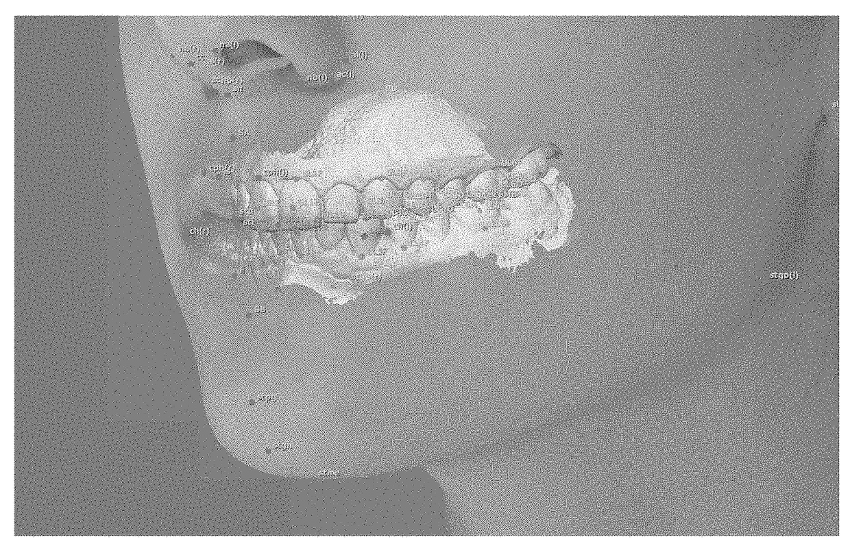
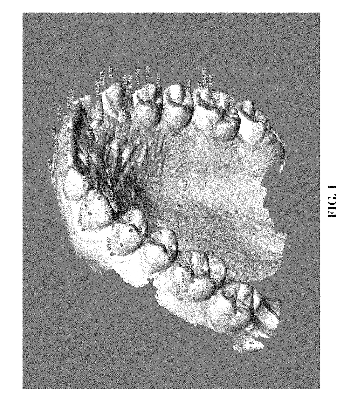
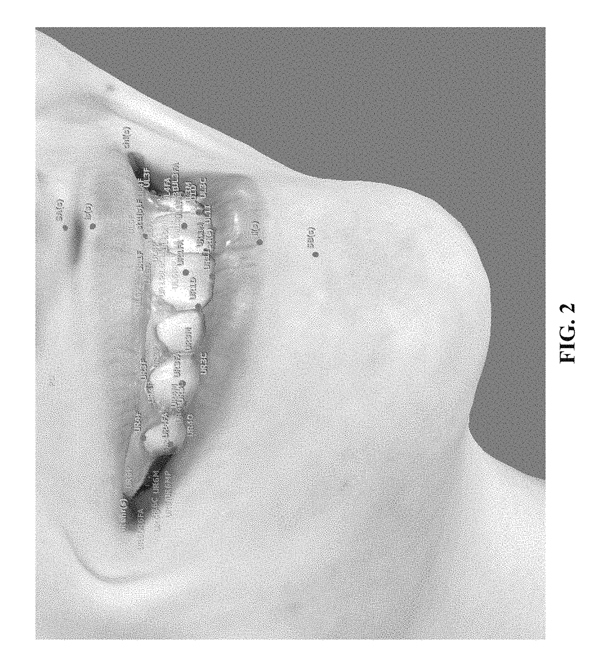
[0078]FIGS. 12, 15 and 16 show the face and teeth of an orthodontic patient with a palatally displaced upper left lateral and incisor buccal segments that are more class II on the left side. Table 8A shows an example of an analysis comparing the patient's values to the reference standard values. The comparison shows that the patient has slightly protrusive upper and lower jaws with average vertical and transverse relationships. For example, the Maxillary Lip Position was 3.7 standard deviations greater than the norm. The upper incisors are two standard deviations greater than the standard and the lower incisors are only one standard deviation more proclined than the standard. This is indicated by Maxillary Incisors (distance and angle) being 2.8 and 2.1 times the standard deviation from the norm, respectively. In accordance with some embodiments of the invention, an orthodontist could seek to treat this condition, for example, by extracting the first upper premolars to reduce the pr...


PUM


Abstract

A reference standard or reference system for dental feature location and orientation can include evaluating the facial appearance of one or more subjects based on aesthetic criteria and selecting a subset of subjects. The reference standard further includes constructing 3D representations, (e.g., a 3D virtual model), of the facial and dental features of the subset of subjects, from photographs of the face and mouth of each subject and determining the location and / or orientation of one or more dental features for each subject. The location and / or orientation values for each subject can be used to produce an average value and a standard deviation that forms the basis for a reference standard as part of reference system for evaluating patients. The method and system further includes constructing 3D representations (e.g., a 3D virtual model) of the facial and dental features of a patient from photographs of the face and mouth of the patient and determining the location and / or orientation of one or more dental features of the patient in order to compare them to the reference standard and develop a treatment plan for the subject based on differences between the patient's measurements and reference standard. The reference standard can use the pupils (e.g., in the natural head position orientation) as a landmark for registration and scaling of the reference standard to the patient under evaluation.

Description

CROSS-REFERENCE TO RELATED APPLICATIONS[0001]This application claims benefit under 35 U.S.C. §119(e) of the U.S. Provisional Application No. 61 / 954,835, filed on Mar. 18, 2014, the contents of which are incorporated herein by reference in their entirety.STATEMENT REGARDING FEDERALLY SPONSORED RESEARCH[0002]Not ApplicableREFERENCE TO MICROFICHE APPENDIX[0003]Not ApplicableBACKGROUND[0004]Technical Field of the Invention[0005]The present invention is directed to methods and systems for evaluating the facial and dental features of a patient for orthodontic diagnosis and treatment. The present invention is directed to methods and systems for establishing a standard for alignment of dental features of a patient and to methods and systems for comparing the facial and dental features of a patient to the standard to assist in developing a treatment plan and evaluating progress during treatment.[0006]Description of the Prior Art[0007]Before Broadbent [1] Introduced cephalometric radiography ...

Claims


Application Information

Patent Timeline
Patent Type & Authority Patents(United States)
IPC IPC(8): G06K9/00A61B5/00A61C7/00A61B34/10
CPCA61C7/002A61B5/0088A61B34/10G06K9/00208G06K9/00281G06K2209/055A61B2034/105A61B2576/02A61C2007/004G16H30/40G06V20/647G06V40/171G06V2201/033
Inventor MASOUD, MOHAMED I.
Owner TRUSTEES OF BOSTON UNIV
Why Eureka
Try Eureka