
Apparatus and method for providing 2D representation of 3D image data representing an anatomical lumen tree structure

An image data and tree structure technology, which is applied in image data processing, 2D image generation, 3D modeling, etc., can solve the problems of negative discovery, detection and processing failure of health status, user uncertainty, and difficulty in inspection, etc.

Active Publication Date: 2008-03-05
KONINKLIJKE PHILIPS ELECTRONICS NV
View PDF0 Cites 0 Cited by
  • Summary
  • Abstract
  • Description
  • Claims
  • Application Information

AI Technical Summary

Problems solved by technology

[0003] This known process suffers from the disadvantage that the pulmonary vascular tree structure is geometrically very complex and usually extends over 100-400 CT slices, which makes the examination very difficult and time-consuming
Therefore, the process requires very specialized skills and exhibits a high degree of user variability as a result of the difficulty in automating the process
[0004] Additionally, existing computer-aided presentation of detection results has the disadvantage of only being able to report the presence of a positive finding, while the user cannot determine whether a negative finding is due to a health condition or a failure of the detection process

Method used


Image

  • Apparatus and method for providing 2D representation of 3D image data representing an anatomical lumen tree structure
  • Apparatus and method for providing 2D representation of 3D image data representing an anatomical lumen tree structure
  • Apparatus and method for providing 2D representation of 3D image data representing an anatomical lumen tree structure

Examples


Embodiment Construction

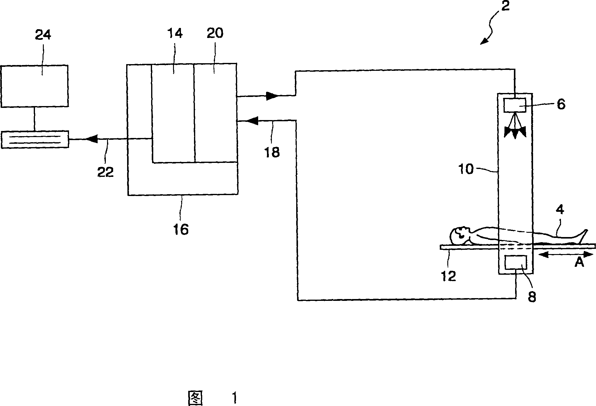
[0049] Referring to FIG. 1 , a computed tomography (CT) scanner apparatus 2 for forming a 3D imaging model of the chest cavity of a patient 4 has an array of X-ray sources 6 and detectors 8 arranged in source / detector pairs around a gantry. 10 in the usual circular arrangement. The setup is shown from the side in Figure 1, so only one source / detector pair can be seen.

[0050] The patient 4 is supported on a platform 12 which is movable in the direction of arrow A in FIG. 1 by suitable means (not shown) under the control of a control unit 14 forming part of a computer 16 . The control unit 14 also controls the operation of the X-ray source 6 and the detector 8 in order to obtain image data of the slice of the patient's body surrounded by the support 10, and the movement of the patient 4 relative to the support 10 is synchronized by the control unit 14 in order to establish a map of the patient's chest cavity. A series of images is typically a stack of 100 to 400 images.

[0...


PUM


Abstract

A CT imaging apparatus (2) for providing a 2D representation of 3D image data representing a pulmonary vessel tree of a patient is disclosed. The apparatus comprises X-ray sources (6) and detectors (8) arranged in pairs for generating 3D image data representing a plurality of positions along lumen branches of the vessel tree and for receiving 3D image data representing at least one anatomical property at each of the locations. A processor (20) processes the 3D image data to provide 2D image data displayed on a display device (24) such that each lumen branch is represented by a line, and the color of each portion of a line representing one of the positions varies in dependence upon the anatomical property being measured.

Description

technical field [0001] The present invention relates to apparatus and methods for providing two-dimensional (2D) representations of three-dimensional (3D) image data representing an anatomical lumen tree structure. The present invention relates particularly, but not exclusively, to apparatus and methods for providing 2D visualization of compressed pulmonary vascular properties. The invention also relates to a computer program product for use in such an arrangement. Background technique [0002] Pulmonary embolism is a life-threatening condition involving blood clotting in the lungs. Pulmonary embolism is known to be detected by high-resolution, contrast-enhanced chest computed tomography (CT) scans. The CT scanner generates image data of transverse slices through the patient's chest, and a 3D model of the patient's pulmonary vascular tree is formed by the CT scanner. The 3D image data is then displayed on a monitor, and the image of the pulmonary vascular tree is inspecte...

Claims


Application Information

Patent Timeline
Patent Type & Authority Applications(China)
IPC IPC(8): G06T17/40
CPCG06T11/206G06T2210/41A61B6/032A61B6/467A61B6/463A61B6/50G06T19/00Y10S128/922
Inventor M·J·奎斯特
Owner KONINKLIJKE PHILIPS ELECTRONICS NV
Learn More