
Efficient user interaction with polygonal meshes for medical image segmentation

A polygon and grid technology, applied in the field of graphics technology, can solve problems such as time-consuming, difficult to confirm the structure of interest in section images, and tediousness, and achieve the effect of fast and accurate planning

Active Publication Date: 2009-07-22
KONINKLIJKE PHILIPS ELECTRONICS NV
View PDF3 Cites 10 Cited by
  • Summary
  • Abstract
  • Description
  • Claims
  • Application Information

AI Technical Summary

Problems solved by technology

This is a time-consuming and tedious process
Also, it is difficult for the user to visually confirm structures of interest in sliced ​​images in order to map attractors
Using Kaus' scheme for such situations requires initial and / or corrective manual adjustments to many of the vertices or nodes of the mesh, which is also a time-consuming and tedious process

Method used


Image

  • Efficient user interaction with polygonal meshes for medical image segmentation
  • Efficient user interaction with polygonal meshes for medical image segmentation
  • Efficient user interaction with polygonal meshes for medical image segmentation

Examples


Embodiment Construction

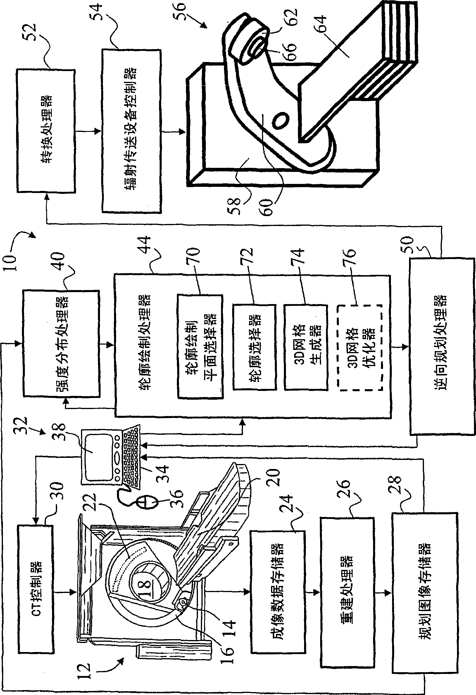
[0027] refer to figure 1, the radiation therapy system 10 includes a computed tomography (CT) scanner 12 for obtaining CT planning images for use in planning an intensity modulated radiation therapy therapy session. CT scanner 12 includes: an x-ray source 14 mounted on a rotating gantry 16 for rotation about an examination area 18; a bed or other support 20 for placing a subject in the examination area 18; and an x-ray detector Array 22 , opposite x-ray source 14 , is arranged on rotating gantry 16 opposite each other for detecting x-rays after they pass through an object in examination region 18 . The CT scanner 12 is used to acquire projection data, such as projection data for multiple axial image slices (such as a multi-slice CT scanner), or a helical three-dimensional projection data set (such as a helical CT scanner), and the like. The projection data is stored in imaging data store 24 and reconstructed by reconstruction processor 26 using a suitable algorithm (eg, filte...


PUM


Abstract

An apparatus for delineating a structure of interest includes: a plane selection interface (32, 70) for selecting a contouring plane of selectable orientation in a three-dimensional image or map (80); a contouring interface (32, 72) for defining a contour in the selected contour plane; and a mesh constructor (74, 76) configured to construct a three-dimensional polygonal mesh (90) delineating the structure of interest in the three-dimensional image or map. The mesh constructor positions constrained vertices (102, Vc) on or near a plurality of non-coplanar delineation contours (84, Ccor, Cobl) defined using the contouring interface.

Description

technical field [0001] The following content deals with graphics technology. The invention is illustrated by way of example for a medical application where structures in a medical image are defined prior to radiation therapy planning or other medical procedure planning. However, the following relates more generally to defining structures in essentially any type of 3D image, 3D map, etc., and to applications other than radiotherapy planning, such as rendering or other post-acquisition image processing, in 3D Features are modeled in the image to enable observation of hidden anatomical structures or other post-acquisition image processing, to estimate absorption properties of structures for subsequent reconstruction of SPECT or PET imaging data, and the like. Background technique [0002] In radiation therapy for tumors (sometimes called radiotherapy), ionizing radiation is used to kill or retard the growth of cancerous tumors or cancerous growth. The problem, however, is tha...

Claims


Application Information

Patent Timeline
Patent Type & Authority Applications(China)
IPC IPC(8): G06T17/20G06T19/20
CPCG06T7/0083G06T2207/30012G06T7/0089G06T2207/30096G06T17/20G06T2207/10072G06T7/12G06T7/149G06T7/0012
Inventor M·R·考斯L·A·道森
Owner KONINKLIJKE PHILIPS ELECTRONICS NV
Features
  • Generate Ideas
  • Intellectual Property
  • Life Sciences
  • Materials
  • Tech Scout
  • Unparalleled Data Quality
  • Higher Quality Content
  • 60% Fewer Hallucinations
Learn More