
Stationary x-ray digital breast tomosynthesis systems and related methods

A tomosynthesis, X-ray technique used in tomosynthesis, patient positioning for diagnosis, instruments for radiological diagnosis, etc.

Active Publication Date: 2010-09-22
THE UNIV OF NORTH CAROLINA AT CHAPEL HILL +1
View PDF1 Cites 51 Cited by
  • Summary
  • Abstract
  • Description
  • Claims
  • Application Information

AI Technical Summary

Problems solved by technology

However, there is currently no efficient way to generate monochromatic or quasi-monochromatic radiation that can provide sufficient X-ray photon flux in a clinical setting

Method used


Image

  • Stationary x-ray digital breast tomosynthesis systems and related methods
  • Stationary x-ray digital breast tomosynthesis systems and related methods
  • Stationary x-ray digital breast tomosynthesis systems and related methods

Examples


Embodiment Construction

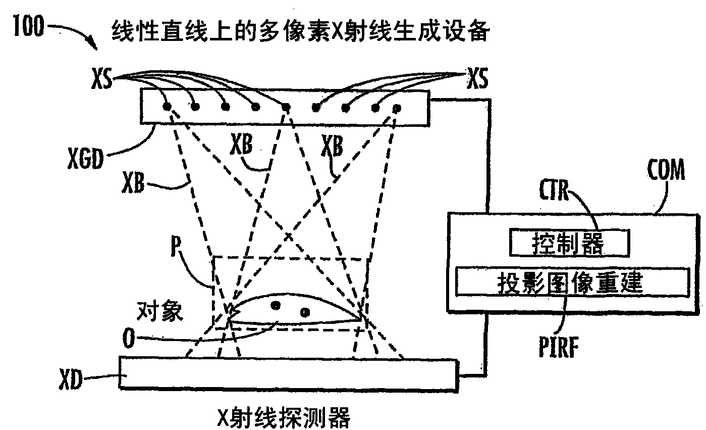
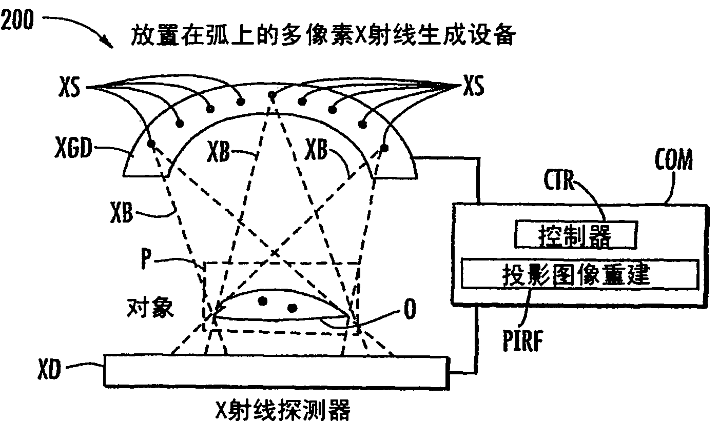
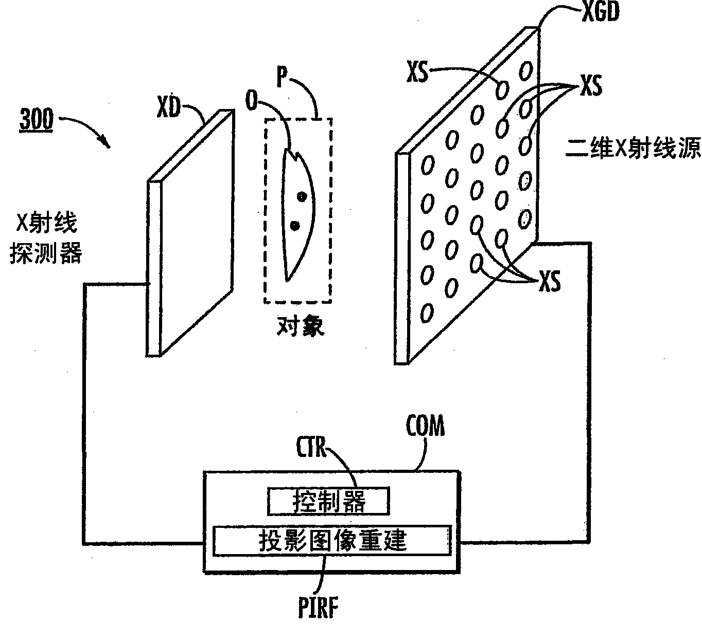
[0033] The subject matter disclosed herein is directed to multi-beam field emission x-ray (MBFEX, also known as multi-pixel field emission x-ray) systems and techniques that can utilize multiple field emission x-ray sources, x-ray detectors, and projection image reconstruction techniques . Specifically, the systems and techniques disclosed herein may be applied to X-ray digital tomosynthesis according to one aspect. According to one embodiment, a plurality of field emission X-ray sources may illuminate a location for locating an object to be imaged with an X-ray beam to generate a projected image of the object. X-ray detectors can detect projected images of objects. The projection image reconstruction function may reconstruct a tomographic image of the object based on the projection image of the object. The subject matter disclosed herein can enable increased scan speed, simplified system design, and enhanced image quality.

[0034] In one application, the subject matter di...


PUM


Abstract

Stationary x-ray digital breast tomosynthesis systems and related methods are disclosed. According to one aspect, the subject matter described herein can include an x-ray tomosynthesis system having a plurality of stationary field emission x-ray sources configured to irradiate a location for positioning an object to be imaged with x-ray beams to generate projection images of the object. An x-ray detector can be configured to detect the projection images of the object. A projection image reconstruction function can be configured to reconstruct tomography images of the object based on the projection images of the object.

Description

[0001] related application [0002] The presently disclosed subject matter claims the benefit of US Provisional Patent Application Serial No. 60 / 961,175, filed July 19, 2007, the disclosure of which is incorporated herein by reference in its entirety. [0003] government interest [0004] The presently disclosed subject matter was made with United States Government support under Grant No. US4CA119343 awarded by the National Cancer Institute. The United States Government therefore has certain rights in the disclosed subject matter. technical field [0005] The subject matter described herein relates to radiography. More specifically, the subject matter describes fixed X-ray digital breast tomosynthesis systems and related methods. Background technique [0006] Mammography is currently the most effective screening and diagnostic tool for early diagnosis of breast cancer and has been responsible for the recent decline in breast cancer mortality. However, the nature of 2D ma...

Claims


Application Information

Patent Timeline
Patent Type & Authority Applications(China)
IPC IPC(8): A61B6/04
CPCA61B6/482A61B6/405A61B6/502A61B6/4291A61B6/025G06T11/006G06T2211/436A61B6/4028A61B6/4007A61B6/584
Inventor O·Z·周杨光卢健平D·拉卢希
Owner THE UNIV OF NORTH CAROLINA AT CHAPEL HILL
Learn More