

Method and system for multi-energy tomosynthesis
a multi-energy tomosynthesis and imaging technology, applied in tomosynthesis, medical science, diagnostics, etc., can solve the problems of limiting their usefulness, reducing its usefulness, and reducing image quality,
- Summary
- Abstract
- Description
- Claims
- Application Information
AI Technical Summary
Benefits of technology
Problems solved by technology
Method used
Image

Examples
Embodiment Construction
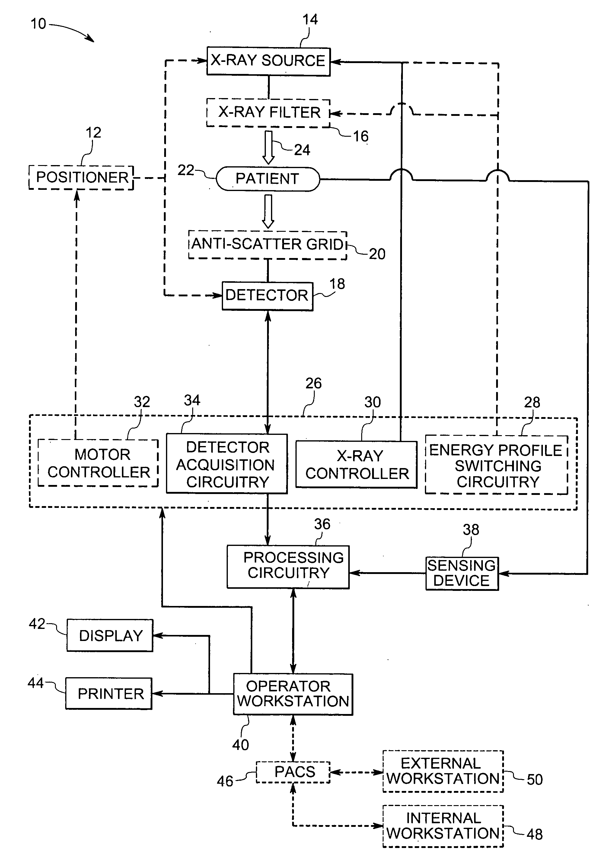
[0018] The present technique is generally directed towards tomosynthesis imaging techniques to generate useful images for medical and non-medical applications. As will be appreciated by those of ordinary skill in the art, the present techniques may be applied in various medical and non-medical applications, such as passenger and / or baggage screening, to provide useful three-dimensional data and context. To facilitate explanation of the present techniques, however, a medical implementation will be generally discussed herein, though it is to be understood that non-medical implementations are also within the scope of the present techniques.
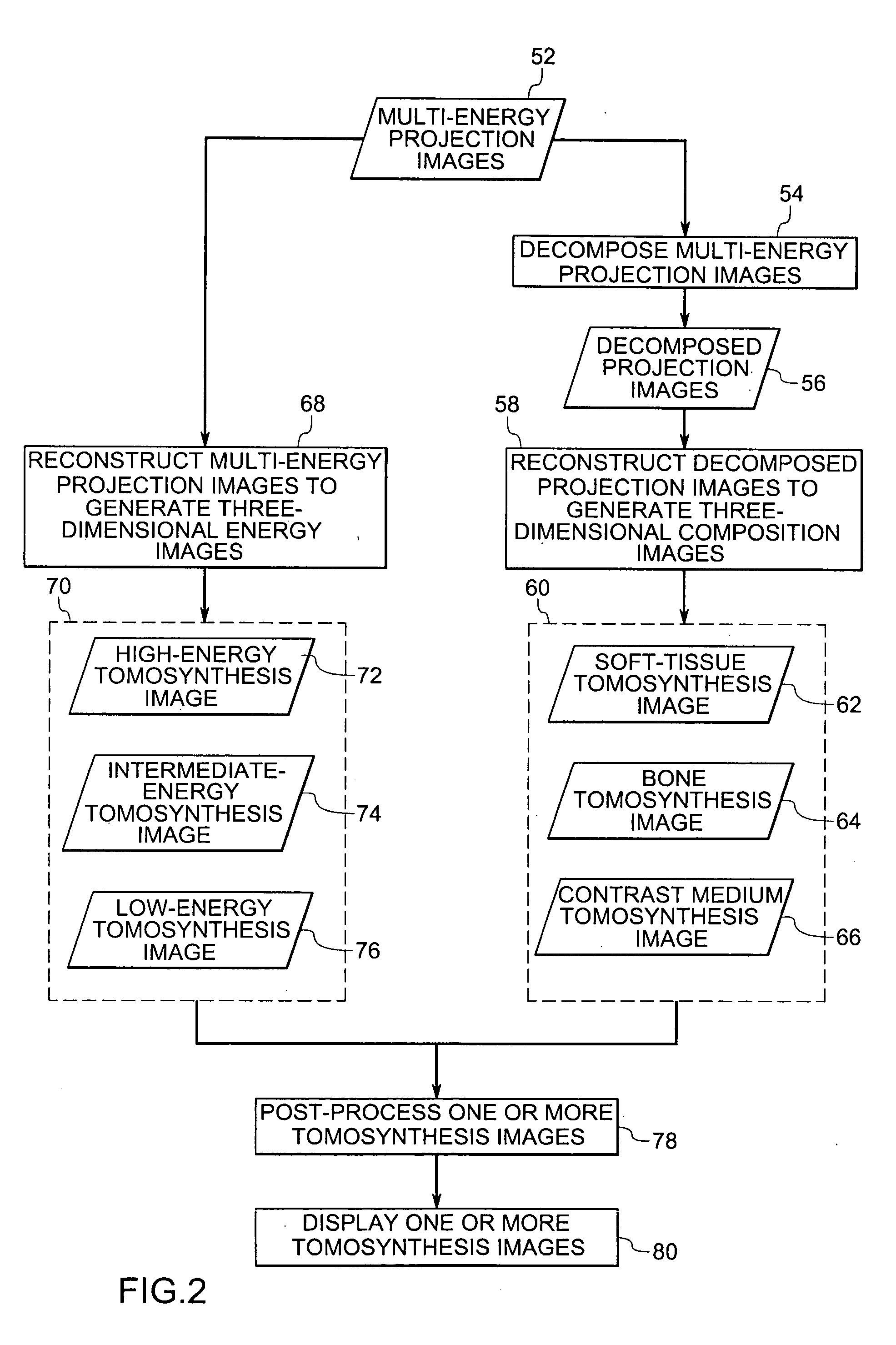
[0019] Tomosynthesis imaging utilizes a limited number of projection images that are acquired over a limited angular range, generally less than 180 degrees, relative to a patient. The projection images are combined and reconstructed to generate three-dimensional images of all or part of the patient. For example, the projection images may be generate...
PUM
 Abstract
Description
Claims
Application Information