Converting low-dose to higher dose 3D tomosynthesis images through machine-learning processes

a machine learning and tomosynthesis technology, applied in image enhancement, instruments, applications, etc., can solve the problems of high breast density, increased cumulative radiation exposure and lifetime attributable risks of radiation-induced breast cancer, and high breast density, so as to achieve less noise, less artifacts, and high quality

Active Publication Date: 2017-03-16
ALARA SYST
View PDF2 Cites 83 Cited by
  • Summary
  • Abstract
  • Description
  • Claims
  • Application Information

AI Technical Summary

Benefits of technology

[0066]This patent specification describes converting lower-quality, lower-dose 3D breast images (or lower-quality, lower-dose 3D tomosynthesis slices or other 3D images) with noise into higher quality, less noise, less artifact, higher-dose-like 3D breast images (or higher-dose-like 3D tomosynthesis slices). The described method and system use a trainable nonlinear regression (TNR) model with a patch-input-pixel-output scheme, which can be called pixel-based TNR (PTNR). A 3D tomosynthesis system acquires multiple projection views (images) of a breast over a limited angle range to reconstruct a 3D volume of high-resolution slices or other 3D images of the breast such as a 3D volume image. In a preferred example, an image patch is extracted from multiple input 2D raw projection views (as a form of images) of a breast acquired at a reduced x-ray radiation dose (lower-dose), and pixel values in the patch are entered into the PTNR as input. The output of the PTNR is a smaller patch, preferably a single pixel, that can correspond to the center pixel of the input image patch. The PTNR receives such patches, for example a respective patch for each pixel of the input image in a raster or some other pattern. The PTNR is trained with matched pairs of raw projection views (images), inputting lower-dose raw projection views (images) together with corresponding desired higher x-ray radiation dose raw projection views (images) (higher-dose views or images), such as standard dose tomosynthesis views or higher-than-standard-dose tomosynthesis views, which are ideal images for the output images. The desired higher-dose x-ray 2D raw projection views may be acquired at up to the highest (or approximately the highest) radiation dose level that the tomosynthesis system can generate or use for practical purposes.
[0067]Through the training, the PTNR learns to convert lower-dose 2D raw projection views (images) to high-dose-like 2D raw projection views (images). Once trained, the trained PTNR does not require the higher-dose tomosynthesis acquisitions anymore. When a new reduced x-ray radiation dose (low dose) 2D raw projection view (image) is entered, the trained PTNR would output a pixel value similar to the corresponding desired pixel value, in other words, it would output high-dose-like 2D raw projection views (images) where noise and artifacts due to low radiation dose are substantially reduced, i.e., views or images with a higher image quality. Note that the new low-dose 2D raw projection views do not have to be acquired at a lower radiation dose level than a standard dose, but they can be acquired at any radiation-dose level lower than the highest radiation dose that the tomosynthesis system can generate or use for practical purposes. Then, from the “high-dose-like” projection views (images), “high-dose-like” 3D tomosynthesis slices (slice images) are reconstructed by using a tomosynthesis reconstruction algorithm [40] such as filtered backprojection [41] and an iterative reconstruction method [42]. The “high-dose-like” 3D tomosynthesis slices where noise and artifact are substantially reduced can be expected to be similar to real high-dose 3D tomosynthesis slices; thus they may be called “virtual” high-dose 3D tomosynthesis slices. With the “virtual high-dose” 3D tomosynthesis slices, the detectability of lesions and clinically important findings such as masses and microcalcifications can be improved. The reconstruction can produce virtual hi-dose other 3D views such as a 3D volume of voxel values for the entire breast or a volume region of interest in the breast, for example by stacking virtual high-dose slices or portions of slices. This patent specification further describes a computer program product that stores in computer-readable media, in non-transitory form, instructions that when loaded into and executed by a computer system carry out the processes described herein.

Problems solved by technology

False positives can lead to “overdiagnosis / overtreatment”.
A significant, if not the main, cause of false-negative results is high breast density that obscures breast tumors in 2D projection mammograms [2].
With breast tomosynthesis, a remaining major challenge in breast cancer screening is radiation exposure.
Repeated breast tomosynthesis for annual screening could increase cumulative radiation exposure and lifetime attributable risks for radiation-induced breast cancer.
Higher radiation doses generally result in higher signal-to-noise ratio, while lower doses generally lead to increased image noise including quantum noise and electronic noise.
Higher radiation exposure and dose would increase the risk of radiation-induced cancer.
Yaffe [23] pointed out, however, that even with use of the 2D synthetic image, radiation dose from 3D breast tomosynthesis can still be an issue, because radiation dose of 3D breast tomosynthesis can be higher than that of 2D mammography.
In addition, because the synthetically created 2D images may appear different from real 2D mammograms that radiologists have been accustomed to, acceptance of the synthetic images by radiologists may still be an issue.
A noise reduction filter may be used, but it tends to smooth out subtle patterns and tiny microcalcifications together with noise.
Thus, it is a challenge to reduce noise and artifact in breast tomosynthesis slices while maintaining diagnostic information and depiction of subtle lesions.

Method used


Image

  • Converting low-dose to higher dose 3D tomosynthesis images through machine-learning processes
  • Converting low-dose to higher dose 3D tomosynthesis images through machine-learning processes
  • Converting low-dose to higher dose 3D tomosynthesis images through machine-learning processes

Examples


Embodiment Construction

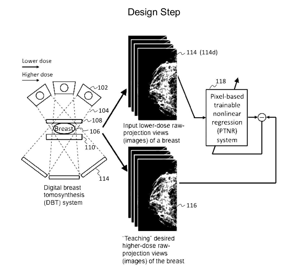
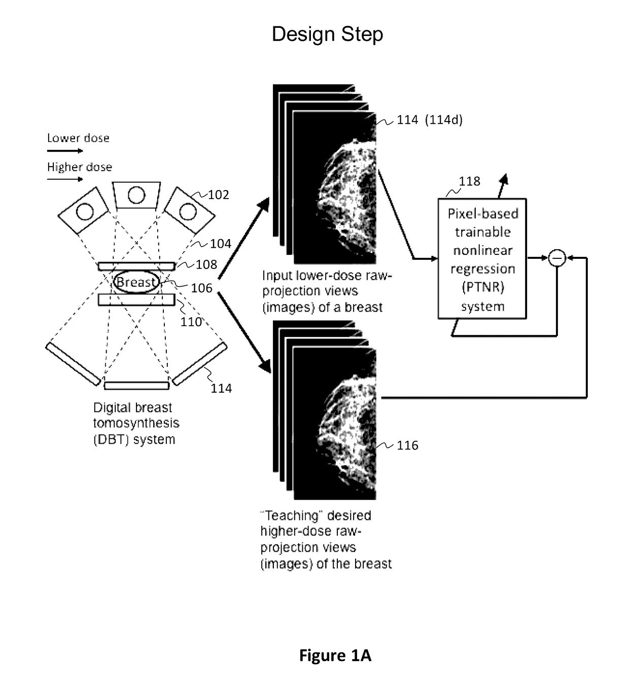
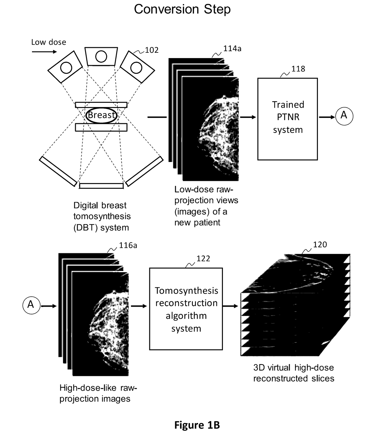
[0077]In preferred examples, the systems and methods described in this patent specification use a pixel-based trainable nonlinear regression (PTNR) that converts lower-dose raw projection views (as a form of images) of a breast to higher-quality, higher-dose-like raw projection views (images) of the breast. Lower-dose raw projection views (images) are of lower image quality, with more noise, than the higher-quality, higher-dose-like raw projection views (images). Higher-dose-like raw projection views (images) look like real, high-dose raw projection views (images) that are of higher image quality with less noise or artifacts than the lower-dose raw projection views (images). The PTNR system uses a trainable nonlinear regression (TNR) model that processes pixels in patches (or regions) in raw projection views (images). There are two main steps associated with PTNR: (1) a design step to determine the parameters in PTNR by using designing pairs of lower-dose (lower image quality) and h...


PUM


Abstract

A method and system for converting low-dose tomosynthesis projection images or reconstructed slices images with noise into higher quality, less noise, higher-dose-like tomosynthesis reconstructed slices, using of a trainable nonlinear regression (TNR) model with a patch-input-pixel-output scheme called a pixel-based TNR (PTNR). An image patch is extracted from an input raw projection views (images) of a breast acquired at a reduced x-ray radiation dose (lower-dose), and pixel values in the patch are entered into the PTNR as input. The output of the PTNR is a single pixel that corresponds to a center pixel of the input image patch. The PTNR is trained with matched pairs of raw projection views (images together with corresponding desired x-ray radiation dose raw projection views (images) (higher-dose). Through the training, the PTNR learns to convert low-dose raw projection images to high-dose-like raw projection images. Once trained, the trained PTNR does not require the higher-dose raw projection images anymore. When a new reduced x-ray radiation dose (low dose) raw projection images is entered, the trained PTNR outputs a pixel value similar to its desired pixel value, in other words, it outputs high-dose-like raw projection images where noise and artifacts due to low radiation dose are substantially reduced, i.e., a higher image quality. Then, from the “high-dose-like” projection views (images), “high-dose-like” 3D tomosynthesis slices are reconstructed by using a tomosynthesis reconstruction algorithm. With the “virtual high-dose” tomosynthesis reconstruction slices, the detectability of lesions and clinically important findings such as masses and microcalcifications can be improved.

Description

REFERENCE TO RELATED APPLICATIONS[0001]This patent application is a continuation-in-part, claims the benefit of, and hereby incorporates by reference parent U.S. patent application Ser. No. 14 / 596,869 filed Jan. 14, 2015 and published as US 2015-0196265 A1, which claims the benefit of U.S. Provisional Application No. 61 / 927,745 filed Jan. 15, 2014.FIELD[0002]This patent specification relates generally to the field of 3D breast imaging, also known as breast tomosynthesis, and more particularly to methods, systems, and computer programs for converting lower-dose tomosynthesis or other breast images into higher-dose-like tomosynthesis or other images.[0003]This patent specification also generally relates to computerized techniques for automated analysis of digital images, for example, as discussed in one or more of U.S. Pat. Nos. 5,751,787; 6,158,888; 7,412,024; 8,378,302; 8,395,120; 6,819,790; 6,754,380; and 7,545,965, and U.S. Publication No. 2006 / 0018524, all of which are hereby inc...

Claims


Application Information

Patent Timeline
Patent Type & Authority Applications(United States)
IPC IPC(8): A61B6/00A61B6/02G06T7/00G06T11/00G06K9/46G06V10/776
CPCA61B6/5205G06T11/005G06K9/4604G06T7/0012G06T2207/30068A61B6/025A61B6/502A61B6/461G06T2207/10116G06T7/0081A61B6/5211A61B6/542G06T2207/20081G06T7/0014G16H50/20G06V10/776G06T5/001G06T5/50G06F18/217
Inventor SUZUKI, KENJI
Owner ALARA SYST
Why Eureka
Try Eureka