Method for visualizing an atrium of the heart in a patient

An atrium, patient technology, applied in the field of visualization of the atrium of the patient's heart, can solve problems such as blood pressure fluctuations

Inactive Publication Date: 2012-04-04
SIEMENS AG
View PDF7 Cites 14 Cited by
  • Summary
  • Abstract
  • Description
  • Claims
  • Application Information

AI Technical Summary

Problems solved by technology

Under the above-mentioned high frequency conditions, not enough blood is pumped from the atrium into the ventricle, and the blood pumped into the blood circulation by the ventricle is also relatively small, resulting in fluctuations in blood pressure

Method used


Image

  • Method for visualizing an atrium of the heart in a patient
  • Method for visualizing an atrium of the heart in a patient
  • Method for visualizing an atrium of the heart in a patient

Examples


Embodiment Construction

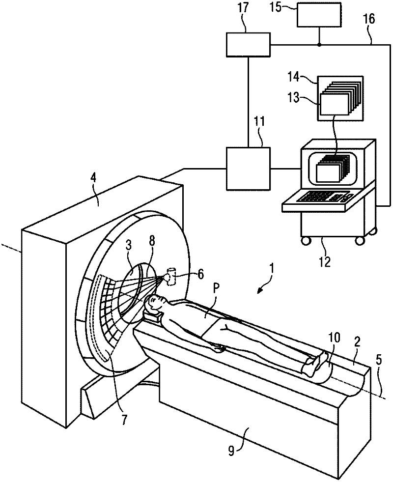
[0027] The same reference signs are used throughout the figures for identical or functionally identical elements, components, structures, etc. The figures shown are schematic only and are not strictly to scale, and the proportions may vary between the figures. The following for figure 1 The shown x-ray computed tomography system 1 is only described in terms of the parts required for understanding the invention without limiting its generality.

[0028] figure 1The computed tomography system 1 shown has a patient couch 2 for supporting a patient P to be examined. The computed tomography system 1 also includes a support 4 which has an x-ray tube-detector system mounted rotatably about a system axis 5 . The x-ray tube-detector system has an x-ray tube 6 and an x-ray detector unit 7 located opposite one another. During operation, X-rays 8 are emitted from the X-ray tube 6 to the X-ray detector unit 7 and collected by the X-ray detector unit 7 .

[0029] The patient couch 2 has...


PUM


Abstract

A method is disclosed for visualizing at least one section of the wall of an atrium (21) of the heart in a patient (P) after an ablation for treating atrial fibrillation, in which, based on a volume data record of the heart in the patient (P) obtained after the ablation, at least the treated atrium (21) of the heart in the patient (P) is segmented, wherein those voxels are established that can be considered part of the inner surface (25), the outer surface (26), and the volume (27) situated between the inner (25) and outer surfaces (26) of the wall of the treated atrium (21). Further, in at least one embodiment of the method, there is volume rendering or ray casting such that only the voxel values of those voxels that lie on the inner surface (25), in the volume (27), or on the outer surface (26) of the wall of the treated atrium (21) are used for visualizing at least the section of the wall of the treated atrium (21) of the heart. At least one embodiment of the invention moreover relates to a computational program (13) that executes the method, a computational unit (12) that carries out the computational program, and / or a data medium (14, 15) with the computational program.

Description

technical field [0001] The present invention relates to a method for visualizing at least a part of the atrial wall of a patient's heart after an ablation procedure for the treatment of atrial fibrillation, so that the result of the ablation procedure can be evaluated. Furthermore, the invention relates to a computer program for implementing the method, a computer unit for executing the computer program, and a data carrier including the computer program. Background technique [0002] Atrial fibrillation is a cardiac arrhythmia in which a rapid succession of chaotic impulses occur from the atrium to the ventricle. The result is that the atria and ventricles contract at different rates independently of each other during atrial fibrillation. Usually the atria and ventricles beat in succession about 70 times per minute. In atrial fibrillation there is a non-directional electrical excitation of the atrium, which leads to a rapid succession of disordered contractions with a freq...

Claims


Application Information

Patent Timeline
Patent Type & Authority Applications(China)
IPC IPC(8): A61B19/00A61B6/03
CPCA61B6/5217A61B6/481G16H50/30
Inventor D.伯恩哈特F.维加-希格拉
Owner SIEMENS AG
Why Eureka
Try Eureka