Horizontal in-situ hybridization detection kit and detection method as well as application for MICRORNA17-3P in earlier stage of cancer pathologic evolution

A detection kit and in situ hybridization technology, applied in the field of related detection technology, can solve the problems of non-decreasing mortality, drug resistance of tumor cells, failure of the anti-cancer war, etc., and achieve high sensitivity, strong specificity, and convenient operation Effect

Inactive Publication Date: 2012-07-04
SUZHOU FUYING GENE TECH
View PDF4 Cites 0 Cited by
  • Summary
  • Abstract
  • Description
  • Claims
  • Application Information

AI Technical Summary

Problems solved by technology

[0003] In 2005, eight institutions including the US Institutes of Health, the Cancer Institute, and the Centers for Disease Control and Prevention made an annual report, reviewing the anti-cancer war launched in 1972. The report believed that human beings had failed in the anti-cancer war, and the conclusion was that cancer died The rate did not decrease, and it listed several factors that caused the failure of the anti-cancer war: 1. Tumor cell heterogeneity (polymorphism); 2. Tumor cell drug resistance; 3. Incomplete design of anti-cancer drugs (animals) unscientific model design), etc.
[0016] In view of the fact that the current clinical diagnosis of cancer (imaging medicine and biochemical indicators are all diagnosed after tumor formation) is a late diagnosis, the treatment is also a late treatment, leading to a treatment model that does not reduce the mortality rate

Method used


Image

  • Horizontal in-situ hybridization detection kit and detection method as well as application for MICRORNA17-3P in earlier stage of cancer pathologic evolution
  • Horizontal in-situ hybridization detection kit and detection method as well as application for MICRORNA17-3P in earlier stage of cancer pathologic evolution
  • Horizontal in-situ hybridization detection kit and detection method as well as application for MICRORNA17-3P in earlier stage of cancer pathologic evolution

Examples


Embodiment 1

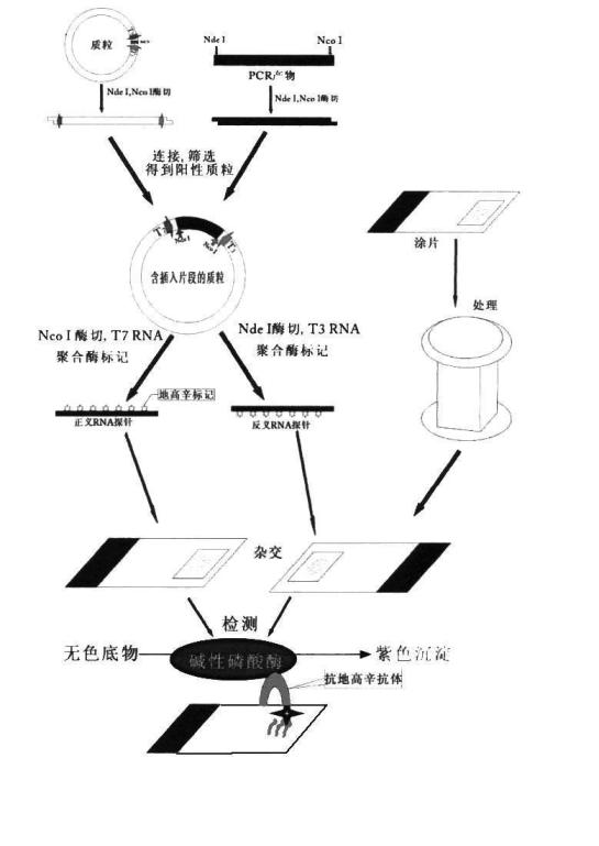
[0051] The in situ hybridization kit of this example was prepared according to conventional methods, and the kit included hybridization probes designed with MICRORNA17-3P, markers, and instructions, wherein: the probe markers of this example were digoxin.

[0052] digestive juice 100μL / tube 1 tube / box colorless transparent liquid protective fluid 100μL / tube 1 tube / box colorless transparent liquid Pre-hybridization solution 1300μL / tube 2 tubes / box colorless transparent liquid Right-sense hybridization solution 10μL / tube 1 tube / box colorless transparent liquid antisense hybridization solution 10μL / tube 1 tube / box colorless transparent liquid blocking solution 1000μL / tube 1 tube / box colorless transparent liquid Alkaline phosphatase antibody 1 μL / tube 1 tube / box colorless transparent liquid Chromogen A 175μL / tube 1 tube / box yellow liquid Chromogen B 320μL / tube 1 tube / box colorless ...

Embodiment 2

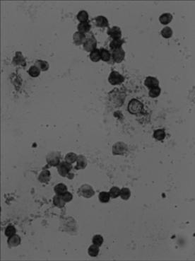
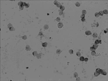
[0060] The implementation process of applying nucleic acid in situ hybridization detection method to the expression level of MICRORNA17-3P in blood samples of each group:

[0061] 1).Take two specimens to be tested;

[0062] 2). Add 50 ml of digestive solution (100 μL of digestive solution plus 99.9 ml of 1× buffer Ⅰ, which is the concentration used) in a glass tank, preheat in a water bath at 37°C for 10 minutes, put 16 slides in, and treat at 37°C for 12 minutes , and then washed with 1× buffer I for 5 min;

[0063] 3). Use 0.2% protection solution (protection solution 1ml plus 1× buffer , 99ml is the concentration used), washed for 10 minutes, washed with three-distilled water for 5 minutes (the above process was carried out in a glass tank), took out the slide, and let it dry naturally;

[0064] 4). Put the slides into a humidifying box, add 25 μL / slice of pre-hybridization solution (add to the place where there are cells), cover with a cover glass, cover the humidifying ...


PUM


Abstract

The invention discloses an in-situ hybridization detection kit which comprises a hybridization probe and a marker. The invention also discloses an in-situ hybridization detection method for microRNA-17-3p being closely related to the early pathologic evolution of various cancers by using the kit. The in-situ hybridization detection method comprises the following steps of: (1) under the condition that the hybridization probe and a target sequence can form a stable hybridization complex, leading RNA (Ribonucleic Acid) to be tested in a substrate to be in contact with the hybridization probe, and forming the hybridization complex; and (2) detecting the hybridization complex. The kit and the detection method provided by the invention can be used for detecting the expression quantity of the microRNA17-3p at the level of RNA, which is more early obtained in comparison with detection indexes of imaging medicine and the traditional clinical biochemistry, thereby the RNA level screening can be realized truly in the earlier stage of canceration, and simultaneously, the detection method provided by the invention is simple and convenient and low in cost and is convenient to popularize and apply in county-level hospitals.

Description

technical field [0001] The present invention relates to the field of biological detection, more specifically, relates to the relevant detection technology related to the change of RNA expression (pathological evolution process) of various cancer pathological evolutions. Background technique [0002] According to the information provided by authoritative organizations at home and abroad, there are 2.6 million new cancer cases in my country every year, nearly 2.1 million deaths, and more than 7 million patients. Globally, there are 8 million new cancer patients every year, nearly 8 million deaths, and more than 8,400 patients. Ten thousand people, the number will double by 2020, this is a set of terrible figures. The cost of cancer diagnosis and treatment is getting higher and higher. The annual treatment cost of cancer patients is 200,000 yuan (may be higher in poor areas, and 200,000 yuan in developed areas). For more than 7 million patients, the annual cost is 1.4 trillion y...

Claims


Application Information

Patent Timeline
Patent Type & Authority Applications(China)
IPC IPC(8): C12Q1/68
Inventor 张云福裘建英裘霖张玉丽
Owner SUZHOU FUYING GENE TECH
Why Eureka
Try Eureka