
Method for reconstructing sparse degree CT (Computed Tomography) image

A sparse-angle, CT image technology, applied in the field of reconstruction of sparse-angle CT images, can solve problems such as poor effect of sparse-angle CT images, long scanning time, blurred stripe artifact structure, etc.

Active Publication Date: 2014-06-04
SOUTHERN MEDICAL UNIVERSITY
View PDF2 Cites 0 Cited by
  • Summary
  • Abstract
  • Description
  • Claims
  • Application Information

AI Technical Summary

Problems solved by technology

Therefore, the current CT used in hospitals still has the following defects under the premise of ensuring image quality: 1. It is necessary to obtain hundreds to nearly a thousand CT projection data and use the filtered back-projection (FBP) reconstruction method to obtain satisfactory results. Therefore, it is inevitable that the radiation dose to the patient is too large; 2. During the scanning time of acquiring nearly a thousand CT projection data, the patient may have body movement, which will also have a certain impact on the subsequent imaging quality, so the Patient scan time needs to be further shortened
Although the method described in the above patent application can reconstruct a high-quality CT image by collecting a large amount of low-dose projection data under the condition of reducing mAs, it still has the following defects: 1. Although the tube current is reduced, nearly a thousand projections need to be acquired data (984 pieces), so the scanning time is relatively long, which will inevitably increase the quality of the reconstructed image caused by the patient's movement; 2. The method described in the patent application with the publication number CN 102314698A cannot be applied to sparse projection CT images Reconstruction, the specific performance is that the reconstructed image is blurred
In the case of collecting a small amount of projection data under normal tube current, due to the small number of projections, the image itself reconstructed by FBP not only contains a lot of noise, but also contains serious streak artifacts and structural blur. The full variational image restoration method in this patent, the full variational image restoration method at this time will take on the difficult task of eliminating noise, artifacts and blurring, it still performs well for denoising, but for deblurring, the effect But it is very poor, because for the total variation deblurring of blurred images, the corresponding convolution matrix must be processed to deblur, and the convolution matrix is ​​usually not easy to obtain, so it can only be "blind" to Blurred and thus poor results
In addition, the image after each iteration of the improved EM reconstruction under a small number of projections contains a large number of blurred components, so the method described in the patent application with the publication number CN 102314698A is not effective in reconstructing sparse-angle CT images.

Method used


Image

  • Method for reconstructing sparse degree CT (Computed Tomography) image
  • Method for reconstructing sparse degree CT (Computed Tomography) image
  • Method for reconstructing sparse degree CT (Computed Tomography) image

Examples


Embodiment Construction

[0035] This embodiment describes in detail the specific implementation process of the reconstruction method of the present invention by taking a sparse-angle CT image of the chest of a lung cancer patient as an example.

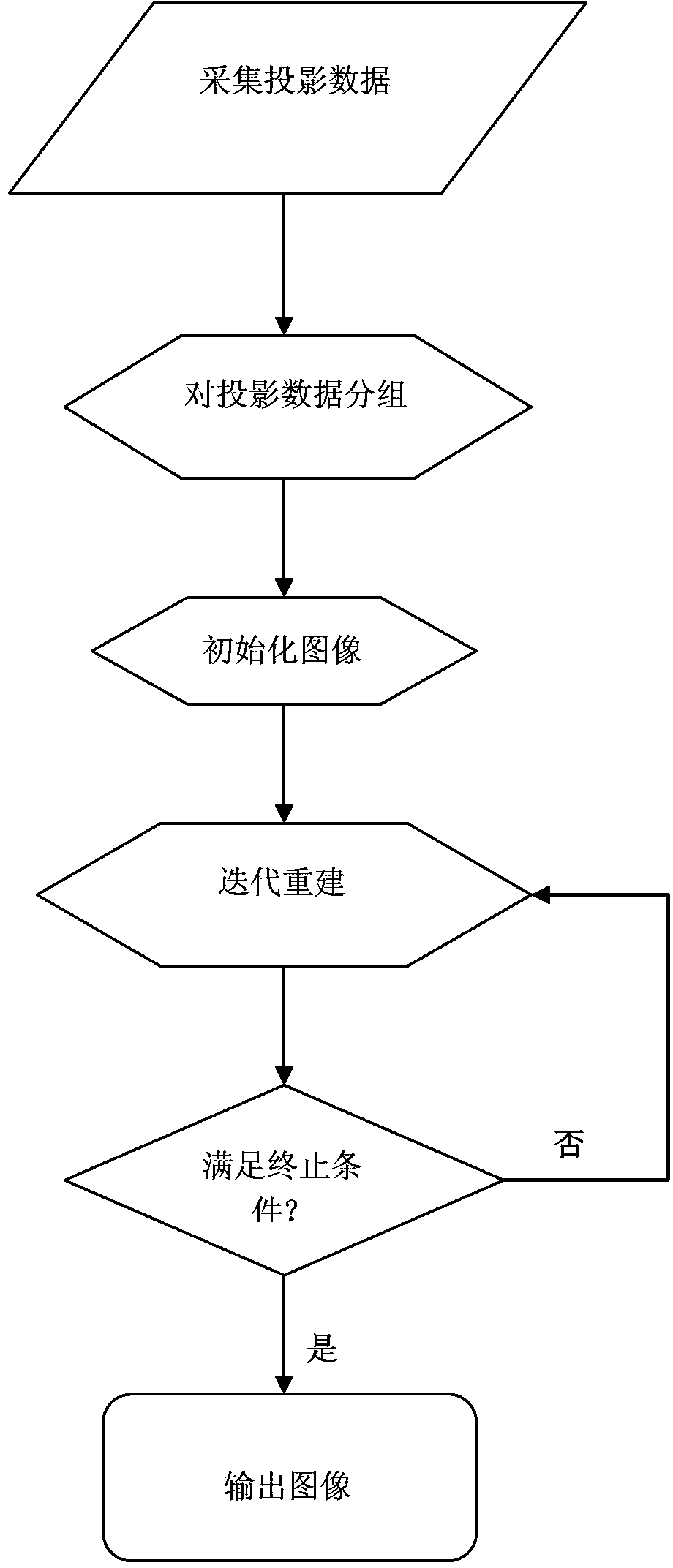
[0036] see figure 1 , the implementation process of this embodiment is as follows.

[0037] 1. Start the GE lightspeed 16-row CT machine, make the tube of the CT machine rotate for one circle, and sequentially collect 72 projection data of lung cancer patients with sparse chest images, and then the projection data of the 72 images collected are divided into 6 groups, each group is the projection data of 12 pictures; record the system parameters of the CT machine at the same time.

[0038] 2. Let α=-1 and β=0.001 in the formula (I) and formula (II) described in the summary of the invention, and use the formula (I) as the reconstruction model to solve the formula (II) obtained by using the auxiliary function method The iterative operation method shown is reco...


PUM


Abstract

The invention relates to an image processing method, in particular to a method for reconstructing a sparse degree CT (Computed Tomography) image. The method comprises the following steps: 1) acquiring a system parameter of a CT machine and sparse degree projection data during a period, and equally dividing the acquired sparse degree projection data into a plurality of groups according to the time sequence; and 2) carrying out cycle iteration operation by using an iterative operation method shown in the formula (II) solved by using the formula (I) as the reconstruction model and using an auxiliary function method until the cycle index reaches the preset value, and using the acquired iteration operation result as the reconstruction image. The reconstruction image obtained by using the reconstruction method has an obvious and clear structure and the contrast ratio of the whole image is remarkably improved.

Description

technical field [0001] The invention relates to image data processing, in particular to a method for reconstructing a sparse-angle CT image. The reconstructed CT image can be used for clinical diagnosis and image-guided radiation therapy. Background technique [0002] Computed tomography, or CT (Computed Tomography), is an essential imaging means to obtain information about the patient's tomographic structure. CT image reconstruction refers to the patient's tomographic information obtained by using a certain reconstruction method from the projection data of the patient collected by the detector. With the rapid development of flat panel detectors, cone beam CT (Cone Beam CT, CBCT) is widely used in clinical diagnosis (such as chest imaging) and image-guided radiation therapy in tumor radiation therapy. Compared with traditional mammography irradiation, the chest volume information reconstructed by CBCT has greatly improved tissue detection accuracy. In image-guided radiatio...

Claims


Application Information

Patent Timeline
Patent Type & Authority Patents(China)
IPC IPC(8): G06T11/00
Inventor 周凌宏齐宏亮徐圆
Owner SOUTHERN MEDICAL UNIVERSITY
Learn More