
Splicing method and system for DR images

A splicing system and image technology, applied in the field of DR image processing, can solve the problems of low efficiency and time-consuming processing, and achieve good visual unity, reduce time consumption, and facilitate medical diagnosis

Inactive Publication Date: 2013-09-11
SHENZHEN LANDWIND IND
View PDF4 Cites 18 Cited by
  • Summary
  • Abstract
  • Description
  • Claims
  • Application Information

AI Technical Summary

Problems solved by technology

This technical solution is effective for ultrasound images, but it uses optimization algorithms such as the least squares method, which consumes processing time
However, since the DR image size is much larger than the ultrasound image, this method is less efficient in processing DR images

Method used


Image

  • Splicing method and system for DR images
  • Splicing method and system for DR images
  • Splicing method and system for DR images

Examples


Embodiment Construction

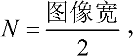
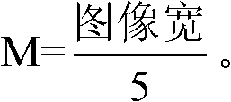
[0025] The present invention down-samples the resolution of the two DR images by reading in the two DR images involved in splicing, and uses the high correlation of the two DR images in the low-resolution image to estimate the displacement between the two DR images. In order to determine the approximate position where the two DR images are aligned, and then finally determine the splicing position of the two DR images through mutual information at the original resolution, and finally adjust the brightness difference of the spliced ​​DR image through grayscale changes, so that the entire spliced DR images achieve visual unity.

[0026] The present invention is suitable for splicing chest radiographs and DR images of limbs, and temporarily does not involve geometric distortion caused by shooting angles.

[0027] The assumption is that the two DR images involved in stitching are adjacent up and down, and the overlapping height is not less than 50 pixels, and the overlapping area i...


PUM


Abstract

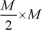
The invention discloses a splicing method and system for DR images. The method comprises selecting an image block of M*M pixels in an expected overlap area of one of the DR images as a reference block, selecting a block with a sliding window with the size of M*M pixels in the expected overlap area of another DR image, calculating the mutual information between each selected block and the reference block, finding out the selected block with the largest mutual information, conforming the splicing position of the two DR images by calculating the coordinate difference of the top left corner vertex of the selected block with the largest mutual information and the top left corner vertex of the reference block, and carrying out splicing processing according to the splicing position after the two DR images are aligned. According to the splicing method and system for the DR images, a multiresolution mode is adopted to carry out image registration, time consumption of image splicing is reduced, the image splicing position is accurately located through the calculation of the mutual information, the spliced image is made to have good vision uniformity by gray level adjustment and seam removing of the image, and medical diagnosis based on the DR images is facilitated.

Description

technical field [0001] The present invention relates to a DR image processing technology, in particular to a DR image splicing method and system. Background technique [0002] With the continuous development of modern medical technology and the continuous improvement of medical diagnosis methods, DR images realized by direct digital X-ray photography system (Digital Radiography, DR) are widely used in clinical diagnosis, medical research and other DR image analysis. [0003] Especially in image-guided orthopedic diagnosis and treatment, it has important clinical application value. At present, the direct digital X-ray imaging equipment (DR) commonly used in orthopedic clinics can only provide images of part of the patient's body. In traditional DR image analysis, doctors need to analyze a series of DR images during diagnosis and treatment, in order to reconstruct a complete patient's whole-body DR image in their minds, which increases the burden on doctors. Therefore, it is...

Claims


Application Information

Patent Timeline
Patent Type & Authority Applications(China)
IPC IPC(8): G06T5/50
Inventor 赵明康雨
Owner SHENZHEN LANDWIND IND
Learn More