Scanning imaging method and system for computed tomography (CT) machine and CT machine

A scanning imaging and gantry technology, applied in computed tomography scanners, echo tomography, etc., can solve problems such as image differences, and achieve the effect of reducing radiation dose and X-ray radiation dose

Active Publication Date: 2013-10-23
SIEMENS SHANGHAI MEDICAL EQUIP LTD
View PDF6 Cites 2 Cited by
  • Summary
  • Abstract
  • Description
  • Claims
  • Application Information

AI Technical Summary

Problems solved by technology

[0011] In practical applications, in order to facilitate image reconstruction, the scanning range of each slice, that is, the field of measurement (Field of Measurement, FOM) is usually larger than the actual region of interest. In t

Method used


Image

  • Scanning imaging method and system for computed tomography (CT) machine and CT machine
  • Scanning imaging method and system for computed tomography (CT) machine and CT machine
  • Scanning imaging method and system for computed tomography (CT) machine and CT machine

Examples


Embodiment Construction

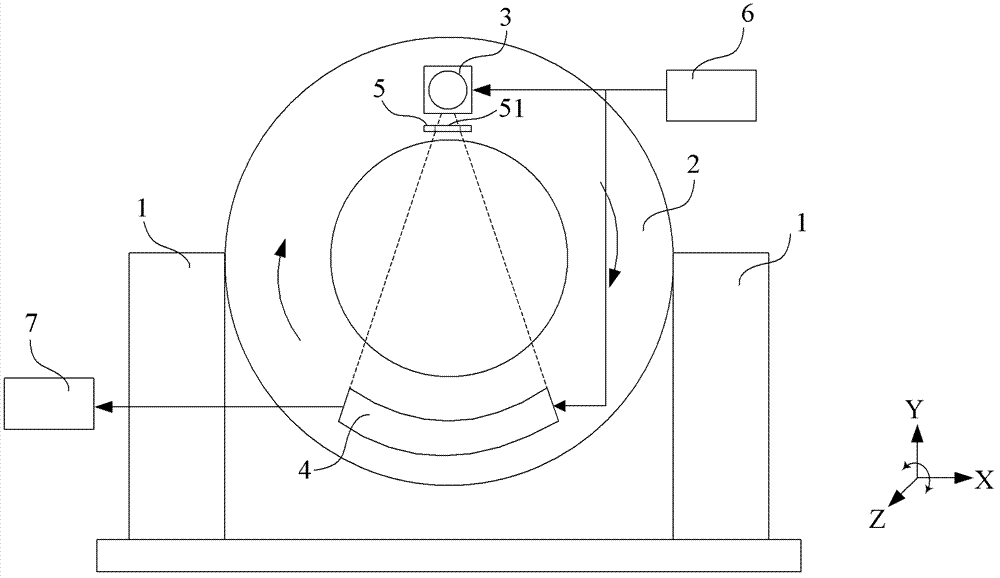
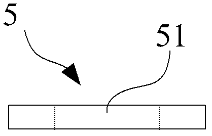
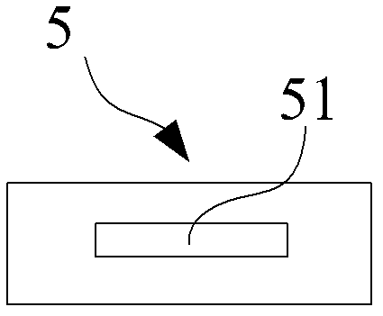
[0051] In order to reduce the total radiation dose of X-rays, considering the characteristics of the reconstructed images between different scans when the same target area is repeatedly scanned, that is, the reconstructed images between different scans may only have image differences in the region of interest, that is, different The images of the non-interest regions of the reconstructed images between scans are almost the same. Consider only using a larger scan range for the initial scan, and use a smaller scan range for subsequent repeat scans. When performing image reconstruction , using the image data obtained from the initial scan to supplement the missing data for the image data obtained by the subsequent scan. Therefore, without affecting the quality of image reconstruction, the X-ray radiation dose for subsequent repeated scans can be reduced, thereby reducing the total radiation dose.

[0052] In order to make the purpose, technical solution and advantages of the pres...


PUM


Abstract

The invention discloses a scanning imaging method and system for a computed tomography (CT) machine and the CT machine. A collimator in the CT machine is provided with a first X-ray constraint hole and a second X-ray constraint hole, and the size of the second X-ray constraint hole in a machine frame rotating tangential direction is smaller than that of the first X-ray constraint hole. The scanning imaging method comprises the steps of utilizing X-rays constrained through the first X-ray constraint hole to perform primary scanning on a target area, and obtaining first image data of the target area; utilizing X-rays constrained through the second X-ray constraint hole to perform current scanning on the target area, and obtaining second image data of the target area; enabling image data in the first image data corresponding to the positions of the second image data to be replaced with the second image data, and obtaining third image data; and reestablishing currently scanned images according to the third image data. According to the technical scheme, the total X-ray radiation dosage can be reduced.

Description

technical field [0001] The invention relates to the field of medical equipment, in particular to a scanning imaging method of a computed tomography (CT) machine, a scanning imaging system of a CT machine and a CT machine. Background technique [0002] As a kind of medical equipment, CT machine is widely used in the medical and health field to assist doctors in imaging patients. figure 1 A structural schematic diagram of a current CT machine is shown in . Such as figure 1 As shown, the CT machine includes: a bottom supporting part 1, a frame (gantry) 2, an X-ray tube 3, an X-ray detector 4, a X-ray tube 3 and an X-ray detector 4 Between the collimator 5, a control device 6 and an image reconstruction device 7. [0003] Wherein, the frame 2 is generally a ring structure, and is installed on the bottom support member 1, and can rotate around its own axis. Usually, the direction parallel to the axis of the rack 2 is called the Z direction, the horizontal direction perpendicu...

Claims


Application Information

Patent Timeline
IPC IPC(8): A61B6/03
Inventor 李涛涛田毅
Owner SIEMENS SHANGHAI MEDICAL EQUIP LTD
Why Eureka
Try Eureka