A method for outputting blood flow parameter images under low radiation dose

A parametric image and output method technology, applied in 2D image generation, image enhancement, image data processing, etc., can solve problems such as large calculation errors and inability to provide more stable blood flow parameters, so as to improve safety and reduce X effect of radiation dose

Inactive Publication Date: 2018-09-28
方若谷
View PDF2 Cites 0 Cited by
  • Summary
  • Abstract
  • Description
  • Claims
  • Application Information

AI Technical Summary

Problems solved by technology

However, these methods are independently calculated for each voxel, so the instability is strong, the calculation error is relatively large, and the context information of the image cannot be effectively used to provide more stable blood flow parameters.

Method used


Image

  • A method for outputting blood flow parameter images under low radiation dose

Examples


Embodiment Construction

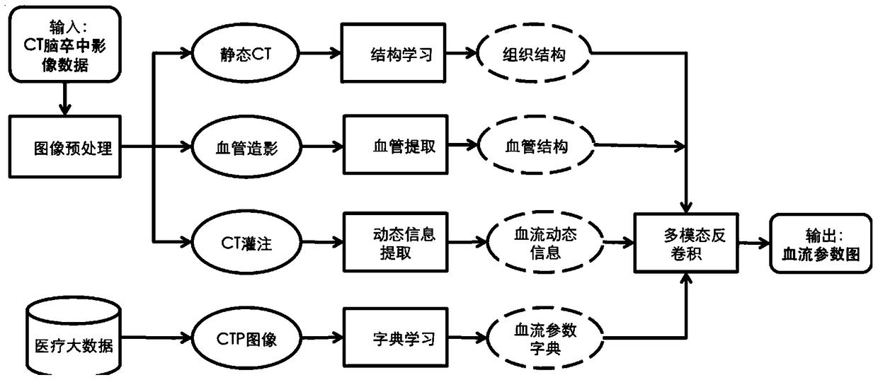
[0027] In the prior art, the technical solution closest to the present invention is: a. Sparse deconvolution method based on learning high-dose hemodynamic parameter images (Fang et al.2013). This method mainly uses dictionary learning and sparse coding methods to learn characteristic dictionaries from a large number of high-radiation dose, high-quality hemodynamic parameter maps, and then uses sparse coding methods to reconstruct image segments in low radiation doses. . The Gai method uses machine learning and data mining methods to learn structural information from existing medical images, which is used to reduce the radiation dose of CTP and improve the accuracy of blood flow parameter maps. (Fang et al.2013) aimed at learning structural information from blood flow parameter maps with high radiation doses of different patients, while ignoring the patient's own non-contrast CT (Non-contrast CT) and CT blood vessels in the stroke diagnostic imaging scheme Angiography (CTAngi...


PUM


Abstract

An output method for blood flow parameter images under a low radiation dose comprises the following steps: acquiring CT (Computed Tomography) cerebral apoplexy image data under the low radiation dose; pre-processing the CT cerebral apoplexy image data; acquiring image characteristics of the pre-processed CT cerebral apoplexy image data; matching the image characteristics in a large medical database to obtain matching results; and outputting the blood flow parameter images according to the matching results. According to the output method provided by the invention, complementary multi-modal medical data is effectively utilized. The radiation dose of X rays accepted by patients in CT perfusion medicinal images is reduced to a great extent, the medical safety is improved and the use value of CT for acute cerebral apoplexy is improved.

Description

technical field [0001] The invention relates to the technical field of computer-implemented medical methods, and specifically relates to a method for obtaining the same high-quality hemodynamic parameter images while reducing the existing radiation dose to 1 / 10. Background technique [0002] CT perfusion is the most commonly used medical imaging method for diagnosis and treatment of cerebrovascular diseases, especially stroke patients. However, in the prior art, continuous brain CT image sampling is performed in order to obtain hemodynamic information. When sampling , the radiation dose of the stroke imaging program including CTP is equivalent to 8-9 times the radiation dose of ordinary chest X-scan or static brain and abdominal CT. Stroke patients need to scan CTP multiple times, and the accumulated radiation dose will destroy healthy cells and increase the risk of cancer. [0003] The study of reducing the radiation dose of CT perfusion has attracted attention since 2000....

Claims


Application Information

Patent Timeline
Patent Type & Authority Patents(China)
IPC IPC(8): G06T11/00G06T5/50
Inventor 方若谷
Owner 方若谷
Why Eureka
Try Eureka