Three-dimensional renal cortex positioning method based on three-dimensional active contour model and three-dimensional Harvard transformation

An active contour model and Haval transform technology, which is applied in the field of medical image processing and analysis, can solve problems such as inability to segment images, and achieve the effect of overcoming low efficiency

Inactive Publication Date: 2014-03-26
SUZHOU UNIV
View PDF1 Cites 5 Cited by
  • Summary
  • Abstract
  • Description
  • Claims
  • Application Information

AI Technical Summary

Problems solved by technology

Although the Harvard transform is fast and reliable, it can on

Method used


Image

  • Three-dimensional renal cortex positioning method based on three-dimensional active contour model and three-dimensional Harvard transformation
  • Three-dimensional renal cortex positioning method based on three-dimensional active contour model and three-dimensional Harvard transformation
  • Three-dimensional renal cortex positioning method based on three-dimensional active contour model and three-dimensional Harvard transformation

Examples


Embodiment Construction

[0046] In order to make the technical means, creative features, goals and effects achieved by the present invention easy to understand, the present invention will be further described below in conjunction with specific embodiments.

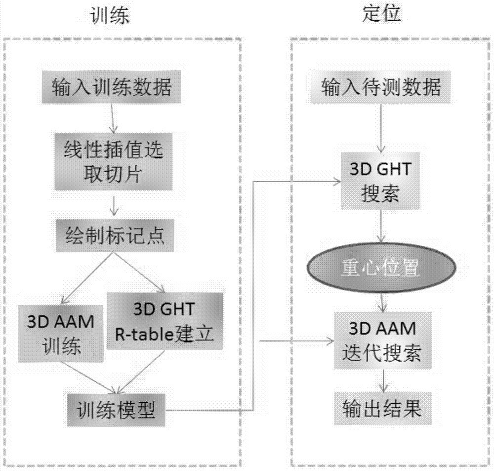
[0047] see figure 1 , the three-dimensional renal cortex positioning method based on the three-dimensional active contour model and the three-dimensional Haval transform of the present invention, including a modeling training part and a positioning part;

[0048] The modeling training part includes the following steps:

[0049] (1-1) Select 15 sets of 3D volume data as training data;

[0050] (1-2) Interpolation calibration selection slice:

[0051] Select all 2D CT images of the entire kidney area from the training data, and select the top and bottom (take a 2D CT image including the entire kidney, select one of the top and one of the bottom, and take 32 from the top to the bottom at the same interval slices) each slice; among the selected slice...


PUM


Abstract

The invention discloses a three-dimensional renal cortex positioning method based on a three-dimensional active contour model and three-dimensional Harvard transformation. The method includes the model establishing training part and the positioning part. The model establishing training part includes the steps that 10-20 sets of three-dimensional data are selected as training data; slices are selected through interpolation correction; marking points are drawn; a model is established through the three-dimensional active contour model, and the model is obtained and comprises an abdominal cavity outer contour, a renal cortex outer surface and a renal cortex inner surface; a kidney contour is stored in an R-table. The positioning part includes the step of searching positioning and the step of positioning the renal cortex; in the step of searching positioning, the kidney contour stored in the R-table is used for positioning the gravity center point of the whole kidney through the three-dimensional Harvard transformation; when the renal cortex is positioned, the model obtained through three-dimensional active contour model establishing is placed in the found gravity center of the kidney, and parameterization is performed and the range is regulated to be smaller so that searching can be performed on the gravity center point till the positioning result is obtained. With the method, the specific position of the renal cortex can be fast and effectively positioned.

Description

technical field [0001] The invention relates to a three-dimensional renal cortex positioning method based on a three-dimensional active contour model and a three-dimensional Haval transform, and belongs to the technical field of medical image processing and analysis. Background technique [0002] The renal cortex is the most important functional structure in the kidney, and it is closely related to most kidney diseases and renal dysfunction. However, in CT or MRI images, the renal cortex is similar to the renal medulla, renal pelvis, and adjacent organs in gray scale. It is difficult to observe with naked eyes and to segment by computer. [0003] Coots and Taylor first proposed the statistical point distribution model (PDM) and used it in 2D image segmentation. Its contribution lies in the invention of a simple and automatic method for learning model parameters from a series of matching points. Then Kelemen established a three-dimensional statistical appearance model by me...

Claims


Application Information

Patent Timeline
IPC IPC(8): G06T17/00G06T7/60
Inventor 陈新建金超向德辉
Owner SUZHOU UNIV
Why Eureka
Try Eureka