System and Method for Combined ECG-Echo for Cardiac Diagnosis

a combined ecg-echo and cardiac diagnosis technology, applied in the field of combined ecg-echo for cardiac diagnosis, can solve the problems of poor contractility, mechanical dysfunction, cardiac dysfunction, etc., and achieve the effect of eliminating potential difficulties and speeding up the acquisition of diagnostic data

Inactive Publication Date: 2010-07-01
UNIV OF VIRGINIA ALUMNI PATENTS FOUND
View PDF33 Cites 95 Cited by
  • Summary
  • Abstract
  • Description
  • Claims
  • Application Information

AI Technical Summary

Benefits of technology

[0011]An aspect of various embodiments of the present invention comprises a device, system, method and computer program method that provides, among other things, the low cost, compact size, ease of use, and simple output format of diagnostic ECG and its electrophysiologic information combined with automated quantitative volume, dimensional, and contractile information available from echo. The present system and related methods described herein eliminate inaccurate analysis of the diagnostic ECG wave forms for chamber size and hypertrophy. The device, system, method and computer program method also alleviates the need for a highly trained technician. Accordingly, these advantages allow the proposed device and related methods to replace nearly all standard electrocardiographs.
[0015]The preferred system also has the ability to simultaneously acquire diagnostic ECG data with diagnostic ultrasound data. Existing methods require two separate patient studies and the clinician must correlate the results, attempting to mentally account for intervening changes in patient condition. The system and method described herein not only speeds the acquisition of diagnostic data, by combining both studies, but also eliminates the potential difficulties of correlating ECG and Echo data acquired at different times. The system also enables diagnosis of more subtle conditions which can only be observed by examining ECG and Echo data from the heart cycle or portion thereof.
[0023]An ultrasound transducer with low profile so as to allow placement for continuous monitoring. The transducer and associated hardware includes numerous aspects that improve the efficiency of the ultrasound data collection and image formation that enable the construction of the low-profile transducer. In one embodiment, the transducer may incorporate A / D conversion circuitry that uses direct inphase and quadrature (IQ) sampling of the received echo signal to reduce the amount of data samples that are required, thereby greatly reducing the complexity of the converter and reducing the heat generated by the circuit. Beamforming circuitry may also be integrated into the transducer and may generate image data points from combinations of rotated versions of the direct sampled IQ samples. Apodization weighting factors may be combined with the required phase rotations. The beamformers may operate to generate c-mode images from samples obtained over an echo time window limited to echoes associated with a desired c-mode image depth. Thus, forming c-mode images also limits the number of samples required to be obtained, further simplifying the data sampling and storage requirements. Still further, multiple image points may be generated in a serial fashion from the data set acquired from a single transmit firing event by re-processing the acquired data set with appropriate phase rotations (to simulate delays).
[0024]The transducer may use an oil-based, as opposed to water-based, couplant so as to be non-drying and thus avoiding the inevitable, and rapid, dryout problem with conventional couplants. Alternatively, the system may use a gel based couplant, like that commonly used on ECG leads, to maintain good coupling with very slow drying. Alternatively the system may use a gel-based couplant connected to a fluid reservoir so that liquid lost to the environment is replaced by liquid from the reservoir.
[0025]A transducer designed with low mass and minimal cabling to maximize positional stability. Such a transducer is particularly useful for continuous cardiac monitoring and for measurement during stress tests.
[0026]An interactive system that guides the placement of the echo transducer. Such interactive system may continuously output measures of image quality, such as mean brightness or image contrast, either audibly or visibly, so as to guide the user in placing the transducer in a more effective location.

Problems solved by technology

Cardiac dysfunction can result from mechanical dysfunction (i.e. poor contractility), electrical dysfunction (i.e. poor conductivity), or complex coupled electrical and mechanical problems.
Only relatively expensive and complex imaging tools, such as echocardiography, are currently able to reliably measure critical dimensions.
Specific dimensions of interest including chamber volumes, wall thickness, and septal thickness can only be indirectly assessed by diagnostic ECG with overall poor predictive value for these measurements.
Historically the diagnostic ECG has achieved wide spread clinical application because of its ease of use and relatively low cost, even though it fails to accurately reflect chamber size and wall thickness.
While the echocardiogram (echo) does directly reflect chamber size and wall thickness, traditional echo systems are costly, bulky, require a highly trained technician, and yield results with high dependence upon operator skill and experience.
Further, by using separate ultrasound imaging and diagnostic ECG systems, it may be difficult for the clinician to correlate mechanical behavior and electrical behavior, undermining diagnosis.
Measurement of the size and shape of the P wave and QRS complexes has been correlated with atrial and ventricular dimension and wall thickness, but the diagnostic accuracy of diagnostic ECG for chamber size and hypertrophy is unacceptably poor.
A major problem with the diagnostic ECG is its unreliability in determining normality or abnormality of chamber size and wall thickness.
Notably, diagnostic ECG systems are relatively low in cost and only limited training is required to obtain high quality recordings.
The transition to 3D imaging is typically accompanied by a reduced frame-rate, somewhat reduced spatial resolution, and an increase in system cost.
Although ultrasound shows increasing potential in cardiac applications, high cost and overwhelming system complexity has greatly limited the scope of application.
While the incorporation of a 3-lead ECG indicates when images were acquired relative to systole and diastole, this simple ECG configuration is inadequate for diagnosing electrical dysfunction or identifying interacting electrical and mechanical problems.

Method used


Image

  • System and Method for Combined ECG-Echo for Cardiac Diagnosis
  • System and Method for Combined ECG-Echo for Cardiac Diagnosis
  • System and Method for Combined ECG-Echo for Cardiac Diagnosis

Examples


example no.1

EXAMPLE NO. 1

[0130]While the ECG is a simple test that is used to diagnose cardiac hypertrophy and enlargement (ventricles and atria) it is frequently wrong. A way to assess cardiac hypertrophy and enlargement is with an echocardiogram. However, echo requires a trained technician and is expensive.

[0131]An attribute of an embodiment of present invention is that the ECG should never again be used to diagnose cardiac hypertrophy and enlargement. It may be used for rate, rhythm, conduction disturbance, infarction, etc., but it should never be called upon to assess cardiac hypertrophy and enlargement.

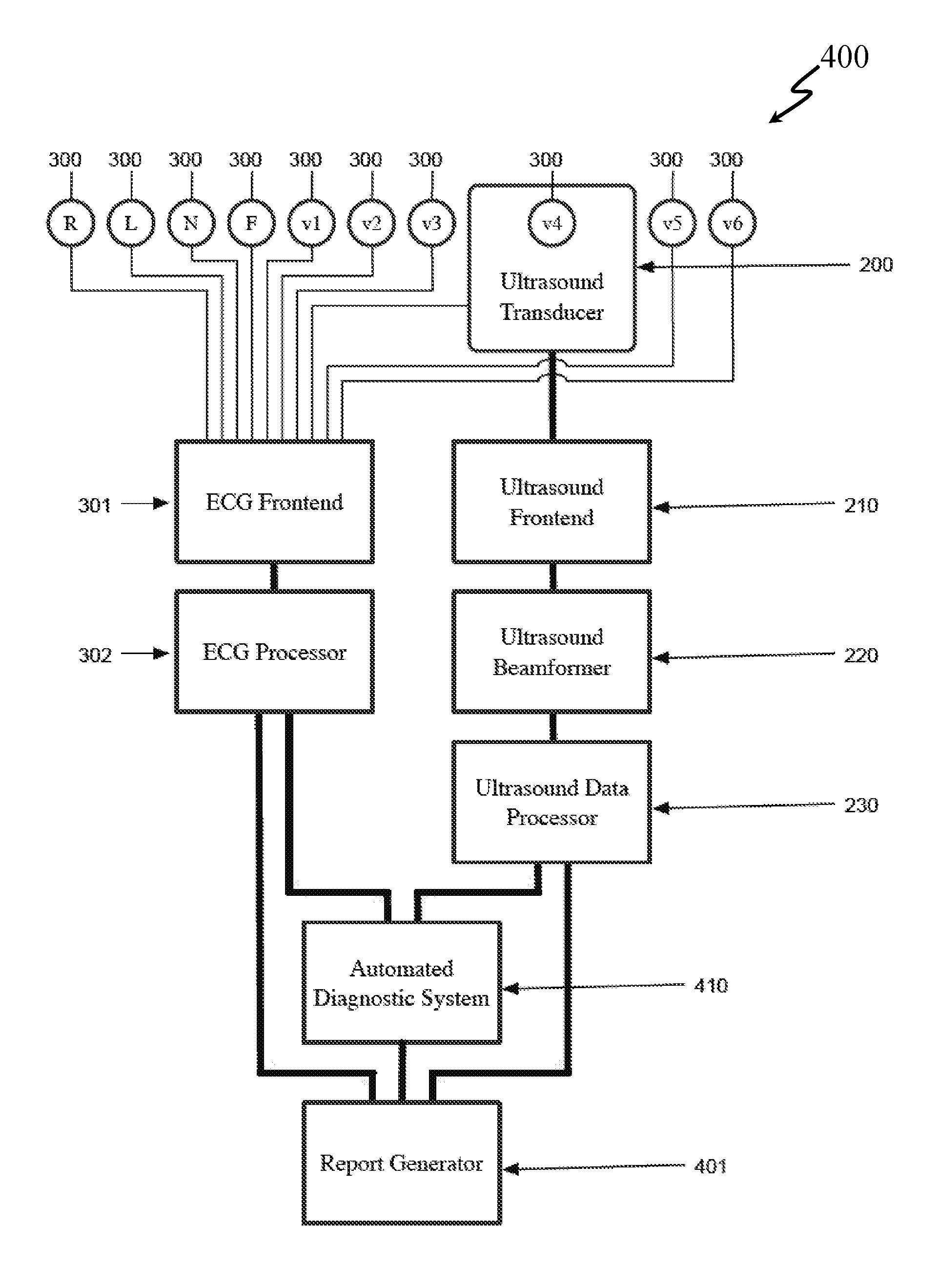
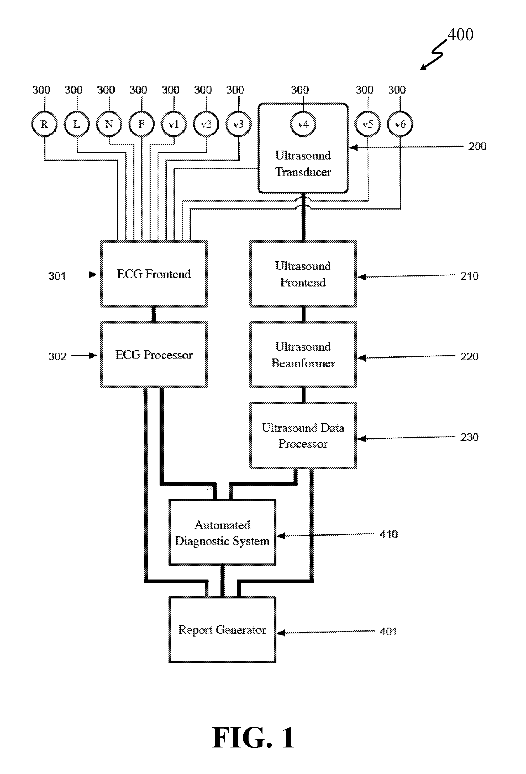
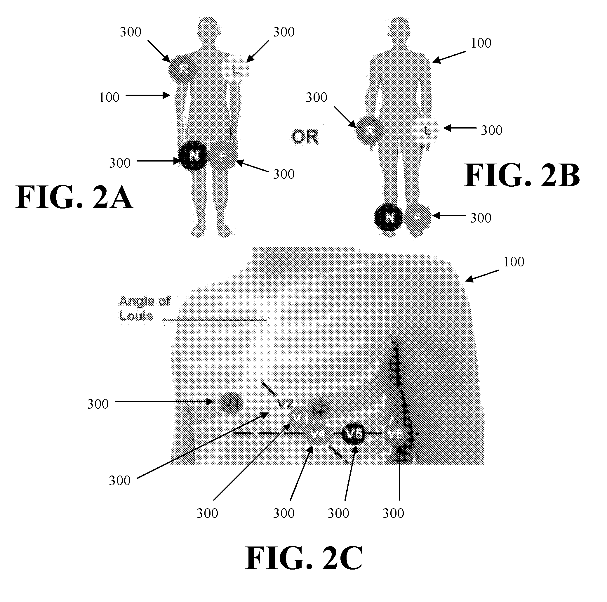
[0132]An approach of an embodiment of the present invention provides an echo transducer and software that automatically measures the size of the heart. A transducer (or small transducer array) would be placed on the chest and a 3-D echo taken, allowing for automated positioning of the image and automated measurement. A technician would not be required. This transducer would be lightweight an...


PUM


Abstract

A system and related method for obtaining volumetric cardiac data of a subject. The data is generated by forming a plurality of focused ultrasound images corresponding to a series of ranges, generating myocardial boundary data for each of the plurality of ultrasound images, calculating the area of the region defined by said myocardial boundary data for each of the plurality of ultrasound images, multiplying the area for each of the plurality of ultrasound images by a slice depth corresponding to said ultrasound image to obtain the slice volume of each slice, and summing the slice volumes to obtain a total volume. In an alternative embodiment the system and related method combine an automated volumetric ultrasound system for finding chamber volumes and myocardial thicknesses, with a diagnostic electrocardiogram system to enable simultaneous diagnosis of mechanical and electrical cardiac problems.

Description

RELATED APPLICATIONS[0001]The present invention claims priority from U.S. Provisional Application Ser. No. 60 / 934,228, filed Jun. 12, 2007, entitled “System and Method for Combined ECG-Echo for Cardiac Diagnosis,” which is hereby incorporated by reference herein in its entirety.GOVERNMENT SUPPORT[0002]Work described herein was supported by Federal Grant Nos. EB002349 and EB001826, awarded by the NIH. The Government has certain rights to the invention.BACKGROUND[0003]The primary function of the heart is as a contractile pump that transports blood to the lungs and throughout the body. While the heart's main function is as a mechanical pump, this pump is driven by an intricate electrical system. Cardiac dysfunction can result from mechanical dysfunction (i.e. poor contractility), electrical dysfunction (i.e. poor conductivity), or complex coupled electrical and mechanical problems. Electrical dysfunction is generally diagnosed clinically using a 12-lead electrocardiogram (ECG). Diagnos...

Claims


Application Information

Patent Timeline
Patent Type & Authority Applications(United States)
IPC IPC(8): A61B8/14A61B5/0402
CPCA61B8/0858A61B8/483A61B8/0883
Inventor GARSON, JR., ARTHURWALKER, WILLIAM F.HOSSACK, JOHN A.BLALOCK, TRAVIS N.
Owner UNIV OF VIRGINIA ALUMNI PATENTS FOUND
Why Eureka
Try Eureka