
A device for free artificial pneumoperitoneum balloon laparoscopy

An inspection device and artificial pneumoperitoneum technology, applied in laparoscopy, endoscopy, medical science, etc., can solve the problems of complex etiology, unclear pain response localization, and high proportion of negative examinations

Inactive Publication Date: 2016-09-28
GENERAL HOSPITAL OF PLA
View PDF9 Cites 0 Cited by
  • Summary
  • Abstract
  • Description
  • Claims
  • Application Information

AI Technical Summary

Problems solved by technology

[0004] (2) General anesthesia is difficult and risky
[0005] (3) The proportion of negative tests is high, making it difficult for patients to accept
Since abdominal organs are innervated by visceral nerves, the localization of pain response is not clear enough, and the etiology of abdominal pain is extremely complex and cumbersome. After exploratory laparotomy or laparoscopy under the premise of huge trauma, no positive or exact lesions were found, so it is difficult to determine It is also difficult to explain the next diagnosis and treatment plan to patients and their families
[0006] The current inspection methods have the above-mentioned disadvantages, the operation difficulty and cost requirements are high, it is difficult to carry out better, and it is even more difficult to provide fast and accurate guidance for disease diagnosis

Method used


Image

  • A device for free artificial pneumoperitoneum balloon laparoscopy
  • A device for free artificial pneumoperitoneum balloon laparoscopy
  • A device for free artificial pneumoperitoneum balloon laparoscopy

Examples


Embodiment Construction

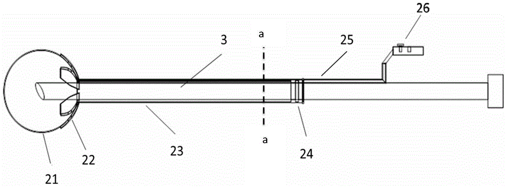
[0026] The invention breaks through the conventional authoritative technology of laparotomy and laparoscopic examination, and provides convenient conditions and technical support for pneumoperitoneum-free abdominal examination. The present invention is described in detail below in conjunction with accompanying drawing:

[0027] see Figure 1A and Figure 1B As shown, the product of the present invention is mainly divided into three parts: the trocar sleeve 1, the balloon inspection device 2 and the inspection endoscope 3, wherein the trocar sleeve 1 and the inspection endoscope 3 parts adopt the existing technology, and Improvement is made on the basis of the existing puncture sleeve, and the end of the original puncture sleeve (the working end of the instrument in contact with the human body is defined as "front" or "front end" in the present invention, such as Figure 1A -1B, Figure 2A and Figure 4 the left end of ; define the control end away from the human body as "rea...


PUM


Abstract

The invention discloses a pneumoperitoneum balloon endoscopy inspection device free of manual operation. The pneumoperitoneum balloon endoscopy inspection device comprises a puncture outfit casing, a balloon inspection device and a check endoscopy which are sleeved in the axial direction from outside to inside, wherein a transparent balloon is formed at the front end of the balloon inspection device; a receptacle support structure which can be folded up is attached to the rear end of the transparent balloon; the rear end of the transparent balloon is connected with a transparent hard connection pipe; a ventilating conduit is embedded in the inside wall of the hard connection pipe; the front end of the ventilating conduit comes into the transparent balloon; and the tail end of the ventilating conduit extends backwards to the rear end of the hard connection pipe and is connected to a one-way vent valve. The device disclosed by the invention can be used under a local anesthesia condition in a non-pneumoperitoneum state; the balloon can effectively create an effective observation space, an abdominal cavity environment and a disease state can be observed favorably, the pneumoperitoneum balloon endoscopy inspection device is mainly applied to abdominal cavity inspection of a disease with unknown etiology in an abdomen, and preoperative assessment staging of patients with abdominal tumors, and a strong reference is provided for diagnosis and treatment in the next step.

Description

technical field [0001] The invention relates to a medical device used in the field of clinical medicine and surgery, which is a device for free artificial pneumoperitoneum balloon endoscopy, in particular a device for free pneumoperitoneum puncture tunnel and balloon endoscopy. Background technique [0002] At present, "exploratory laparotomy" or "exploratory laparoscopy" is routinely required for unexplained abdominal pain or difficult-to-diagnose diseases and tumor stages. The above two methods are both invasive examinations, and they need to be performed under general anesthesia, which leads to the following problems: [0003] (1) Severe trauma: Exploratory laparotomy generally requires a median abdominal incision, or a paramedian incision, or an incision through the rectus abdominis muscle, so that it can be extended up and down or expanded laterally when needed during the operation. The incision is often more than 20 cm. Routine laparoscopy will be due to the CO used i...

Claims


Application Information

Patent Timeline
Patent Type & Authority Patents(China)
IPC IPC(8): A61B1/012A61B1/313
Inventor 陈凛崔建新卢灿荣卫勃王宁郗洪庆
Owner GENERAL HOSPITAL OF PLA
Learn More