Cancer screening method

a screening method and cancer technology, applied in the field of cancer screening methods, can solve the problems of high false negative rate, high cost of manpower, and many undesired properties of pap smear, and achieve the effect of facilitating the diagnosis of other abnormal specimens and accurate screening results

Inactive Publication Date: 2008-12-18
NAT DEFENSE MEDICAL CENT
View PDF0 Cites 116 Cited by
  • Summary
  • Abstract
  • Description
  • Claims
  • Application Information

AI Technical Summary

Benefits of technology

[0011]The invention provides a cancer diagnostic method. The method uses the degree of methylation of a specific gene as the index to diagnose whether there is presence of cancer. The cancer diagnostic method according to the invention is applicable on the detection of cervical cancer. In addition to be the first line screening for cervical cancer, the cancer diagnostic method according to the invention can be used as the second line screening for cervical cancer in combination with or as an assistant to HPV testing in order to achieve a more accurate screening result for cervical cancer. Furthermore, the cancer diagnostic method according to the invention is capable of detecting other cancer types such as ovarian cancer, liver cancer and the like, to facilitate the diagnosis of other abnormal specimens.

Problems solved by technology

There are, however, many undesired properties of Pap smear.
For one, it requires sampling by a physician, and analysis by a medical examiner / pathologist, which is a high cost of manpower that poses difficulty on promoting the test in many developing countries.
Also, Pap smear has a high false negative rate which delays diagnosis and proper treatment prior to cancerous pathological change.
As for HPV testing, although it is highly sensitive, it tends to create a high false positive rate, which not only leaves patients worry in vain, but also wastes much medical resources in examinations follow up to those false positive patients.
However, HPV infection is necessary but not sufficient to cause cervical cancer.
Persistence of HPV infection and viral load may be detrimental accounting the development of HSIL and cancer (Ylitalo, Sorensen et al.
However, the molecular mechanism of cervical carcinogenesis remains illusive.
What genes are specifically methylated in cervical cancer and how many genes are required to achieve clinical application will remain a blossoming issue in the coming future.

Method used


Image

  • Cancer screening method
  • Cancer screening method
  • Cancer screening method

Examples


example 1

Materials and Methods

1. Tissue Specimens

[0066]Cervical tissue specimens were obtained from patients with normal uterine cervixes (n=45) and patients with LSIL (n=45), HSIL (n=58), and invasive squamous cell carcinoma (SCC; n=109) of the uterine cervix. The patients were diagnosed, treated, and tissue banked at the Tri-Service General Hospital, Taipei, Taiwan, since 1993. For diagnostic purposes, cytological, histological, and clinical data for all patients were reviewed by a panel of colposcopists, cytologists, and pathologists. All patients were examined and treated using a standard hospital protocol for cervical neoplasia. Controls were recruited from healthy women who underwent routine Pap screening during the same period. Informed consent was obtained from all patients and control subjects. Exclusion criteria included pregnancy, chronic or acute viral infection, a history of cervical neoplasia, skin or genital warts, an immune-compromised state, the presence of other cancers, an...

example 2

Identification of Methylated Genes in Invasive Squamous Cell Carcinoma of the Cervix

[0077]Differential methylation hybridization (DMH) was carried out by means of CpG island microarrays to screen out the highly methylated gene in cervical squamous cell carcinoma (SCC). The result from CpG island microarrays revealed that there were 216 points exhibited differential methylation between cervical cancer tissue specimens and normal cervical smears, of which, after taking off those having overlapped sequences, 26 gene promoter domain CpG islands (promoter CGIs).

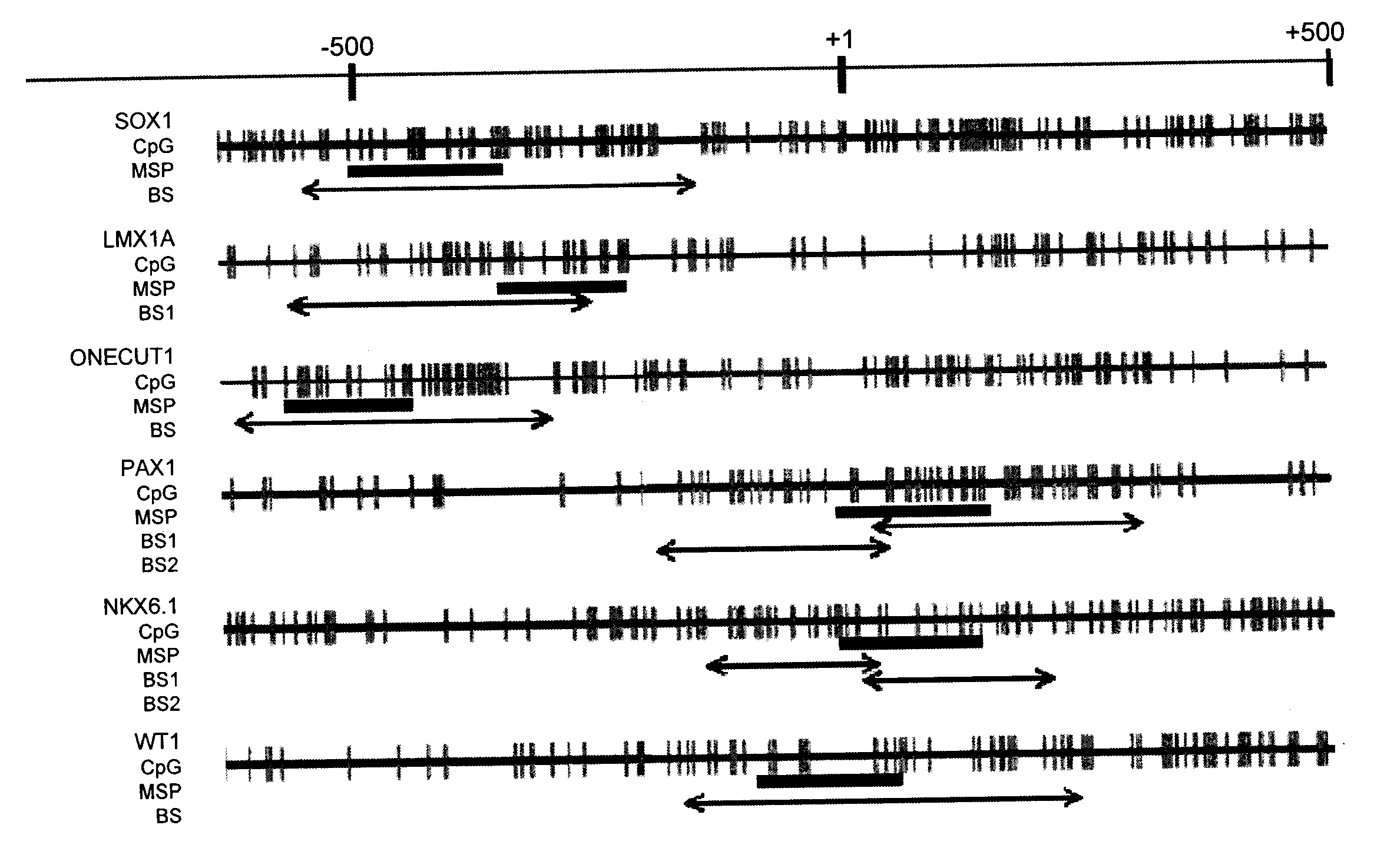
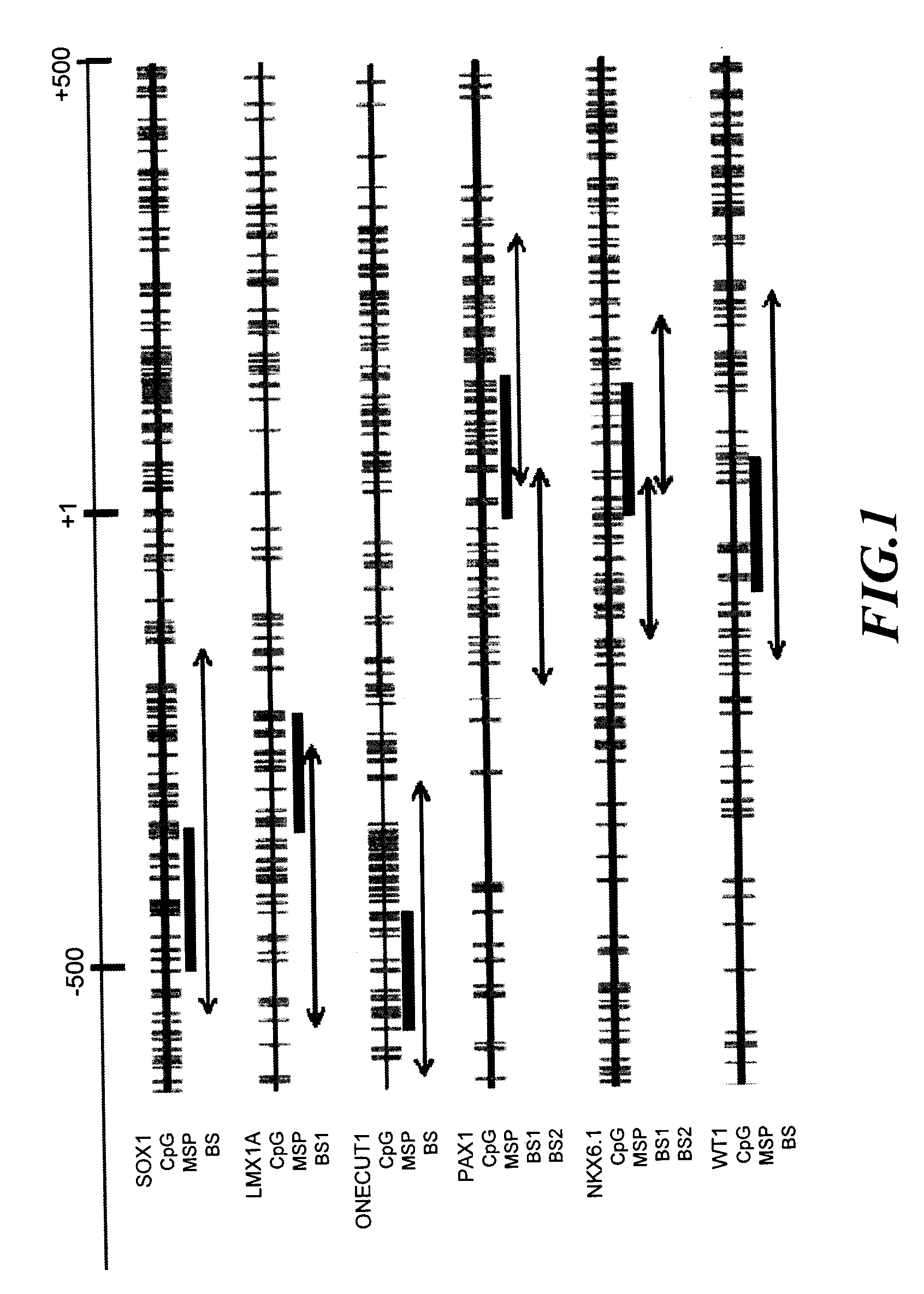
[0078]Sequencing and analysis were carried out on these gene promoter and 6 genes were selected. These genes included: SOX1 (SEQ ID No: 1), PAX1 (SEQ ID No: 2), LMX1A (SEQ ID No: 3), NKX6-1 (SEQ ID No: 4), WT1 (SEQ ID No: 5) and ONECUT1 (SEQ ID No: 6). Their detailed information were shown in Table 4. All of these 6 genes are important transcription factors in the development course, of which, SOX1, PAX1, LMX1A, NKX6-1, and WT1 we...

example 3

Association of DNA Methylation and Gene Expression in HeLa Cervical Cancer Cell Line

[0081]In order to confirm whether the expression of cervical cancer methylation indicator gene is regulated through DNA methylation, HeLa cervical cancer cell line was treated with 10 μM of DNA methyltransferase inhibitor, 5′-aza-2′-deoxycytidine (AZC) (Sigma Chemical Co.), for 4 days, following by checking the demethylation by the 6 gene promoters described above by means of methylation-specific PCR (MSP) carried out with MSP primer (U) that could recognize specifically non-methylated gene sequence, as well as with MSP primer (M) that could recognize specifically methylated gene sequence, respectively. Results as shown in FIG. 4A indicated that among non-5′-aza-2′-deoxycytidine (AZC)-treated HeLa cervical cancer cell lines (AZC−), 6 gene promoters exhibited methylated conditions (as shown at column 1 in FIG. 4A), and no non-methylated gene was detected (as shown at column 2 in FIG. 4A). On the other...


PUM

PropertyMeasurementUnit
volumeaaaaaaaaaa
volumeaaaaaaaaaa
temperatureaaaaaaaaaa

Abstract

A method for screening cancer comprises the following steps: (1) providing a test specimen; (2) detecting the methylation state of the CpG sequence in at least one target gene within the genomic DNA of the test specimen, wherein the target genes is consisted of SOX1, PAX1, LMX1A, NKX6-1, WT1 and ONECUT1; and (3) determining whether there is cancer or cancerous pathological change in the specimen based on the presence or absence of the methylation state in the target gene; wherein method for detecting methylation state is methylation-specific PCR (MSP), quantitative methylation-specific PCR (QMSP), bisulfite sequencing (BS), microarrays, mass spectrometer, denaturing high-performance liquid chromatography (DHPLC), and pyrosequencing.

Description

BACKGROUND OF THE INVENTION[0001]1. Field of the Invention[0002]The invention relates to a cancer screening method, and in particular, to a cancer screening method using methylated DNA as the biomarker.[0003]2. Description of the Prior Art[0004]Cervical cancer has been one of the main causes of death in females worldwide and in Taiwan. Based on the statistical survey by the World Health Organization (WHO) in 2002, cervical cancer was the second major disease responsible for the death of women worldwide, second to breast cancer. Regular cervical cancer screening is the best way to prevent cervical cancer. Conventional cervical cancer screening includes two approaches: the most commonly used Pap smear, and human papilloma virus testing (HPV testing). Pap smear consists of sampling secreta from cervix uteri, examining under microscope whether there is cancerous pathological change in the exfoliated epithelial cell, so as to detect cervical cancer early. HPV testing, on the other hand, ...

Claims


Application Information

Patent Timeline
Patent Type & Authority Applications(United States)
IPC IPC(8): C12Q1/68
CPCC12Q1/6886C12Q2600/112C12Q2600/154C12Q2600/156C12Q2600/16
Inventor LAI, HUNG-CHENG
Owner NAT DEFENSE MEDICAL CENT
Why Eureka
Try Eureka