A Fast Registration and Stitching Method for 3D Digital Subtraction Angiography Images

A subtraction angiography, three-dimensional digital technology, applied in the field of medical image processing, can solve the problems of uneven magnetic field gradient, long processing time, low image signal-to-noise ratio, etc., and achieve the effect of shortening processing time and avoiding influence

Active Publication Date: 2017-11-28
SHANGHAI UNITED IMAGING HEALTHCARE
View PDF2 Cites 0 Cited by
  • Summary
  • Abstract
  • Description
  • Claims
  • Application Information

AI Technical Summary

Problems solved by technology

For multi-segment 3D angiographic subtraction volume data images, since the foreground image of each slice in the vascular subtraction volume data (that is, the highlighted blood vessel with contrast agent) has little information, it is more noise-polluted. Black background, and the black background often contains residual information of bone and soft tissue that has not been completely reduced, making the image signal-to-noise ratio very low. Such image data quality places extremely high requirements on the accuracy of the 3D image registration algorithm, and often The number of slices of vascular subtraction volume data is large, so the registration of 3D images is relatively time-consuming, which eventually leads to a longer processing time for stitching
In addition, since the overlapping areas are at the upper and lower edges of the image, the uneven magnetic field gradient often leads to large information differences in the overlapping areas, which will affect the accuracy of the registration algorithm of the 3D image

Method used


Image

  • A Fast Registration and Stitching Method for 3D Digital Subtraction Angiography Images
  • A Fast Registration and Stitching Method for 3D Digital Subtraction Angiography Images
  • A Fast Registration and Stitching Method for 3D Digital Subtraction Angiography Images

Examples


Embodiment Construction

[0018] The present invention will be further described below in conjunction with the accompanying drawings and embodiments.

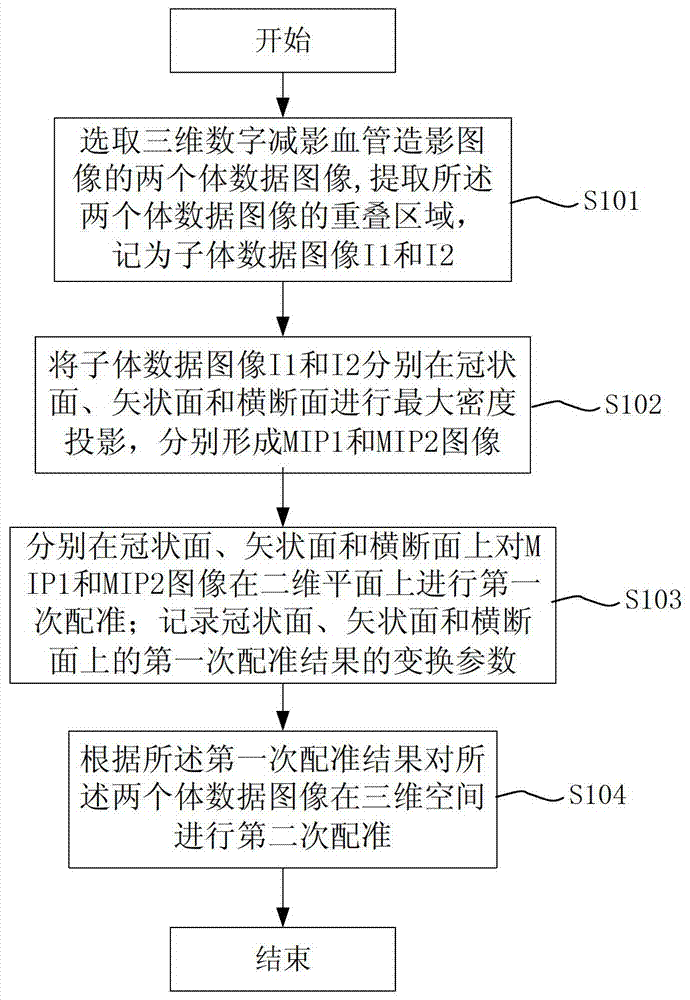
[0019] figure 1 It is a schematic diagram of the rapid registration process for three-dimensional digital subtraction angiography images of the present invention.

[0020] See figure 1 , the rapid registration method for three-dimensional digital subtraction angiography image provided by the present invention comprises the following steps:

[0021] S101: Select the three-dimensional vessel subtraction volume data images of two adjacent blood vessels to cut and segment along the overlapping area, extract the overlapping area of ​​the two volume data images, and obtain two sub-volume data images, which are denoted as sub-volume data images I1 and I2; The three-dimensional vascular subtraction volume data image is an image generated by MR angiography, CT angiography or other imaging methods. Taking the processing of 3D subtraction volume data images by ...


PUM


Abstract

The invention discloses a method for fast registration and mosaic of three-dimensional digital subtraction angiography images, comprising the following steps: a) selecting two volume data images of three-dimensional digital subtraction angiography images, extracting overlapping regions and recording them as sub-volumes Data image; b) Maximum density projection of daughter volume data images on coronal plane, sagittal plane and transverse plane respectively to form MIP1 and MIP2 images; c) First matching of MIP1 and MIP2 images on the above two-dimensional plane respectively d) performing a second registration on the two volume data images in three-dimensional space according to the first registration result, then fusing the overlapping areas, and finally splicing to form a panoramic three-dimensional subtraction volume data image. The rapid registration and mosaic method for three-dimensional digital subtraction angiography images provided by the present invention can avoid the influence of image data quality on the registration accuracy, and greatly shorten the processing time of registration and mosaic.

Description

technical field [0001] The invention relates to a medical image processing method, in particular to a fast registration and mosaic method for three-dimensional digital subtraction angiography images. Background technique [0002] Angiography is currently the main method for diagnosing vascular diseases. In the process of medical imaging, there is often a contradiction that the inspected part is much larger than the area of ​​the image detector. It is necessary to divide the part into several shots, and then stitch them together according to certain rules. Taking magnetic resonance imaging as an example, due to the limitation of the size of the scanning plate of the MRI system, when scanning the blood vessels of the lower extremities or the whole body, the MRI will perform multi-segment scanning, reconstruction and subtraction, in order to synthesize multiple segmented vascular subtraction volume data For a panoramic vascular subtraction volume data, stitching has become nec...

Claims


Application Information

Patent Timeline
Patent Type & Authority Patents(China)
IPC IPC(8): G06T5/50G06T3/40
Inventor 于文君其他发明人请求不公开姓名
Owner SHANGHAI UNITED IMAGING HEALTHCARE
Why Eureka
Try Eureka