Ultrasonic Guidance Of A Needle Path During Biopsy

A path, ultrasound image technology, applied in the field of medical diagnostic imaging systems, can solve problems that hinder the efficiency of biopsy procedures, increase patient discomfort and symptoms, and make it difficult to interpret needle pathways

Active Publication Date: 2014-09-17
KONINKLJIJKE PHILIPS NV
View PDF9 Cites 7 Cited by
  • Summary
  • Abstract
  • Description
  • Claims
  • Application Information

AI Technical Summary

Problems solved by technology

As a result, the efficiency of the biopsy procedure is hampered by the often difficult interpretation of the needle passage, especially for deep needle insertions that may require multiple reinsertions
All the aforementioned difficulties result in additional time for multiple reinsertions of the biopsy needle, thereby increasing patient discomfort and morbidity

Method used


Image

  • Ultrasonic Guidance Of A Needle Path During Biopsy
  • Ultrasonic Guidance Of A Needle Path During Biopsy
  • Ultrasonic Guidance Of A Needle Path During Biopsy

Examples


Embodiment Construction

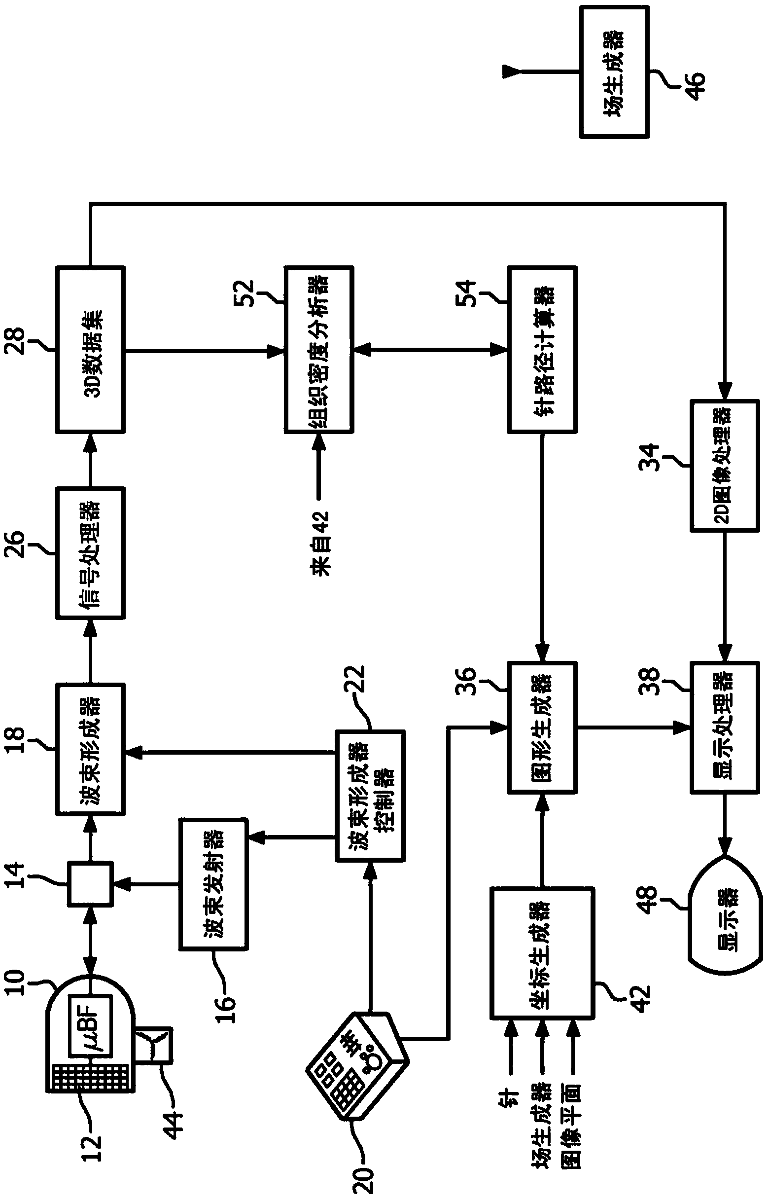
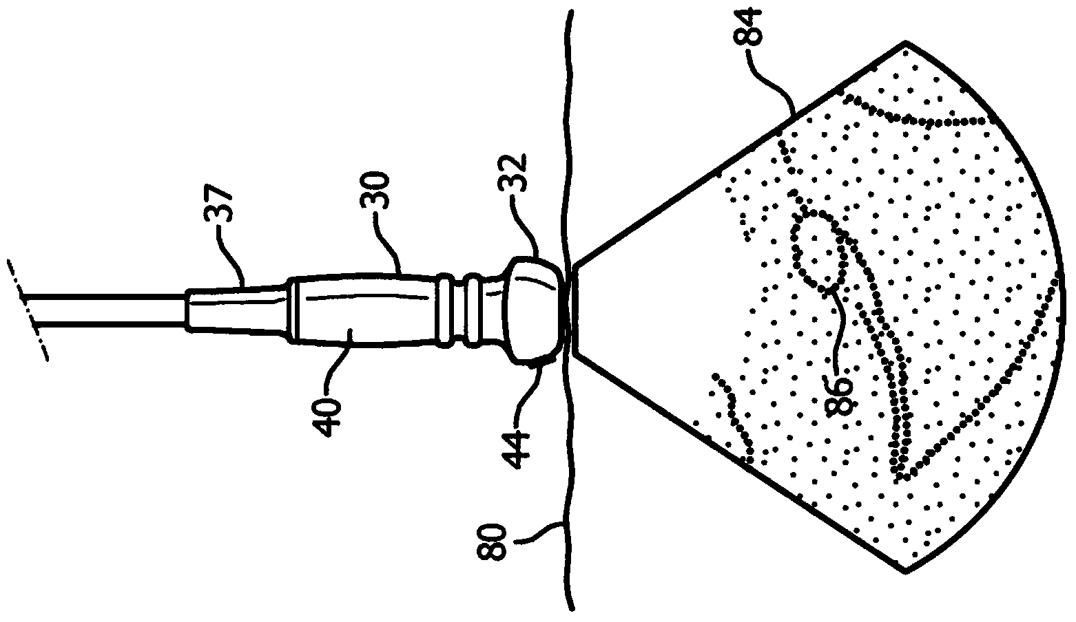
[0013] first reference figure 1 , showing in block diagram form an ultrasound imaging system for assisting needle guidance. The ultrasound probe 10 includes an array transducer 12 to scan and image an area in front of the transducer. The transducer array can be a one-dimensional (1D) array to scan a plane in front of the probe, but preferably the transducer is a two-dimensional (2D) array transducer 12 that emits electrons over a volumetric area The beams are steered and focused, and a single or multiple receive beams are received in response to each transmitted beam. With the 2D array 12, the probe is able to scan the image plane and tissue on either elevational side of the image plane. Groups of contiguous transducer elements in the array (referred to as "slices" or "sub-arrays") are collectively operated by a microbeamformer (μBF) in the probe 12, which performs the processing of the received Partial beamforming of echo signals and thereby reducing the number of conducto...


PUM


Abstract

An ultrasonic imaging system is described which visually assists biopsy needle insertion. The system produces pixel values of pixels along one or more paths of needle insertion. A tissue density analyzer is responsive to the pixel values to estimate tissue density along a path of needle insertion. A needle path calculator is responsive to the tissue density analyzer and proposes one or more favorable paths through imaged tissue for insertion of the biopsy needle. The system may be used in conjunction with a three dimensional navigation system which spatially locates the needle and ultrasound image plane in 3D space.

Description

technical field [0001] The present invention relates to medical diagnostic imaging systems, and more particularly to diagnostic ultrasound systems that provide visual guidance of needles during biopsies or other invasive procedures. Background technique [0002] A biopsy is a medical procedure that involves the removal of cells or tissue for examination. The procedure involves the medical removal of tissue from a living subject to determine the presence or extent of disease. The resected tissue is typically examined under a microscope by a pathologist, and can also be analyzed chemically for the disease state. When the entire mass or suspicious area is removed, the procedure is called an excisional biopsy. When only a sample of tissue is removed, while preserving the histological architecture of the tissue's cells, the procedure is called an incision biopsy or core biopsy. When a needle is used to remove a sample of tissue or fluid in such a way that the cells are excised...

Claims


Application Information

Patent Timeline
Patent Type & Authority Applications(China)
IPC IPC(8): A61B8/08A61B8/00A61B19/00G06T7/00
CPCA61B8/463A61B10/0233A61B2019/5276A61B8/0858A61B8/0841A61B8/483A61B19/201A61B8/5207A61B2019/507A61B90/11A61B2034/107A61B2090/378
Inventor S·R·库达维利R·S·班达卢
Owner KONINKLJIJKE PHILIPS NV
Why Eureka
Try Eureka