Handheld auxiliary scanning device

An auxiliary device and a handheld technology, applied in medical science, acoustic wave diagnosis, infrasonic wave diagnosis, etc., can solve the problems of difficult reconstruction, variability, and limited image angle, etc., and achieve the best effect of ultrasonic imaging quality

Inactive Publication Date: 2015-01-21
安华亿能医疗影像科技(北京)有限公司
View PDF10 Cites 6 Cited by
  • Summary
  • Abstract
  • Description
  • Claims
  • Application Information

AI Technical Summary

Problems solved by technology

[0003] 1. Conventional ultrasound images are two-dimensional, and the diagnostician must mentally transform multiple two-dimensional images to form a complex three-dimensional structure, which leads to variability and sometimes incorrect diagnoses;
[0004] 2. It is very difficult to localize the 2D ultrasound image plane of the organ and reconstruct it afterwards
This makes the approach error-prone and

Method used


Image

  • Handheld auxiliary scanning device
  • Handheld auxiliary scanning device
  • Handheld auxiliary scanning device

Examples


Embodiment Construction

[0049] In order to make the object, technical solution and advantages of the present invention clearer, the present invention will be described in further detail below in conjunction with specific embodiments and with reference to the accompanying drawings.

[0050] It should be noted that all expressions using "first" and "second" in the embodiments of the present invention are to distinguish two entities with the same name but different parameters or parameters that are not the same, see "first" and "second" It is only for the convenience of expression, and should not be construed as a limitation on the embodiments of the present invention, which will not be described one by one in the subsequent embodiments.

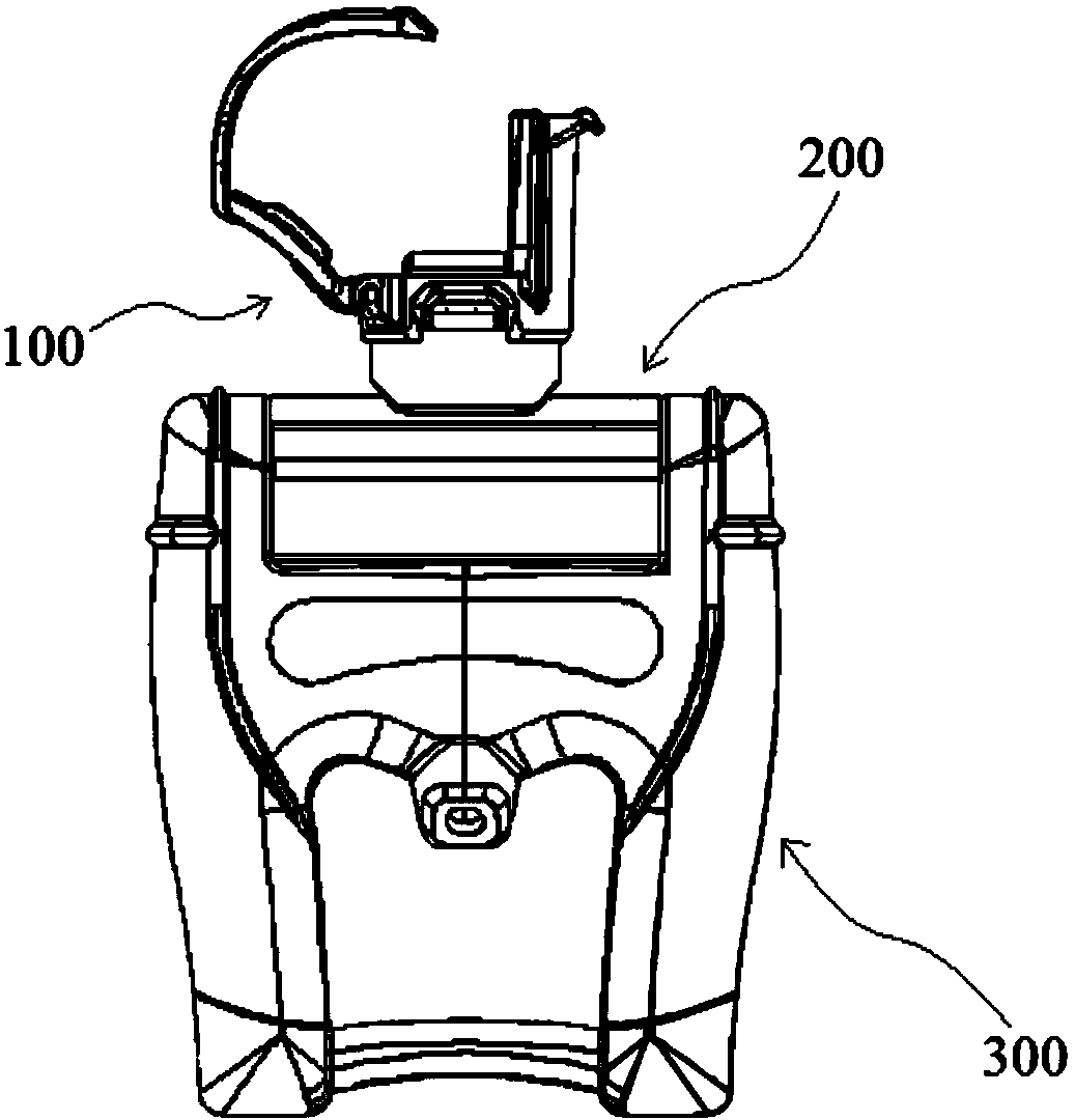
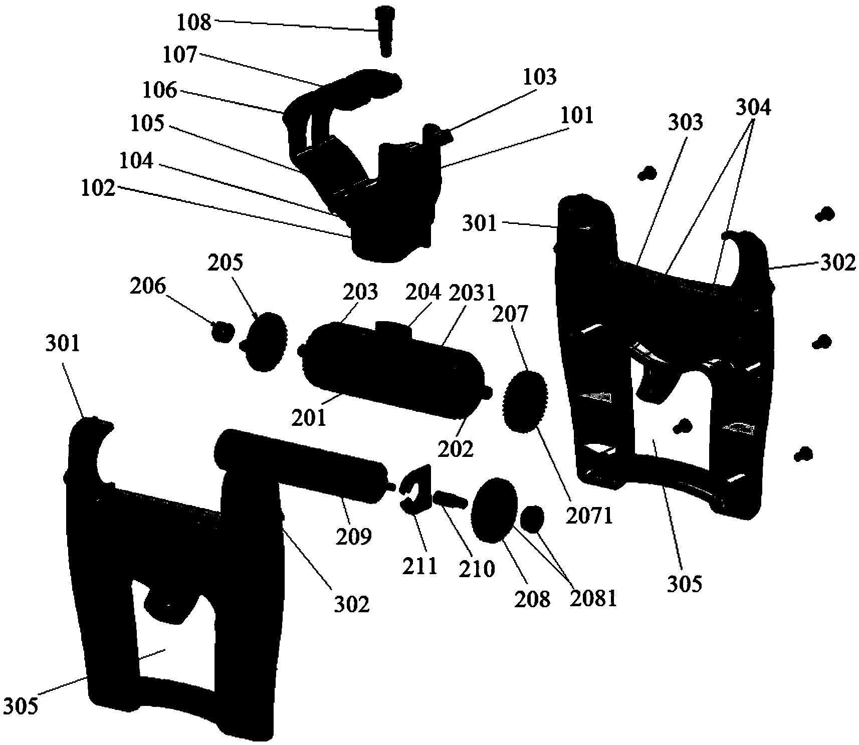
[0051] Refer to attached figure 1 And attached figure 2 , are respectively a structural schematic diagram and an exploded structural schematic diagram of an embodiment of the handheld scanning auxiliary device provided by the present invention.

[0052] The handhel...


PUM


Abstract

The invention discloses a handheld auxiliary scanning device. The handheld auxiliary scanning device comprises an H-shaped hollow shell, a scanning transmission mechanism fixed to the hollow shell, and an ultrasonic probe support in transmission connection with the scanning transmission mechanism. A handheld handle is formed by the two lower ends of the H-shaped hollow shell, an ultrasonic probe is fixed to the ultrasonic probe support, and the scanning transmission mechanism drives the ultrasonic probe support to move in a linear mode so as to enable the ultrasonic probe to continuously and stably collect two-dimensional carotid artery section images at different positions. When used in cooperation with the ultrasonic probe, the handheld auxiliary scanning device can assist the ultrasonic probe to continuously, accurately and stably collect the two-dimensional ultrasonic images.

Description

technical field [0001] The invention relates to the technical field of image acquisition, in particular to a hand-held scanning auxiliary device. Background technique [0002] Medical ultrasound imaging technology is today's mainstream method for easy and non-invasive assessment of the detailed internal structure of tissues and organs for assessment, diagnosis, designation of treatment options, and determination of curative effect. However, conventional 2D ultrasound image scanning has several drawbacks: [0003] 1. Conventional ultrasound images are two-dimensional, and the diagnostician must mentally transform multiple two-dimensional images to form a complex three-dimensional structure, which leads to variability and sometimes incorrect diagnoses; [0004] 2. It is very difficult to localize the 2D ultrasound image plane of the organ and reconstruct it afterwards. This makes the approach error-prone and limited in its ability to quantify disease progression and decline,...

Claims


Application Information

Patent Timeline
IPC IPC(8): A61B8/00
CPCA61B8/4209A61B8/4461A61B8/483
Inventor 汪洋魏桑迪
Owner 安华亿能医疗影像科技(北京)有限公司
Why Eureka
Try Eureka