Culture medium for culturing human amniotic epithelial cells

A technology of epithelial cells and culture medium, which is applied in the biological field to achieve the effects of simple culture, growth inhibition and growth promotion

Active Publication Date: 2015-02-25
吉林和泽生物科技有限公司
View PDF4 Cites 6 Cited by
  • Summary
  • Abstract
  • Description
  • Claims
  • Application Information

AI Technical Summary

Benefits of technology

This patented technology allows us to create cellular layers on surfaces that are used by humans or animals (like babies). These layered structures help them reproduce more effectively when they come into contact with their environment instead of just having three types of cells like those found within our own tissues called placenta. By growing these specialized cells onto specific areas inside the animal's abdomen, we aim to improve its ability to regenerate damaged organs such as heart muscle due to damage caused from diseases.

Problems solved by technology

The technical problem addressed in this patented text relates to studying how humans have developed different types of cell structures called Acenes that help regulate their development during gestational periods. These include neural crest cells found within the basement layer covering the surface of the uterine cervix, neuroectodes located at both ends of the spinal cord, melanocytes located near blood vessels, keratinizing squamous junctions between nerve endings, and other specialized areas such as lymphatic channels and muscle bundling around joints.

Method used


Image

  • Culture medium for culturing human amniotic epithelial cells
  • Culture medium for culturing human amniotic epithelial cells
  • Culture medium for culturing human amniotic epithelial cells

Examples


Embodiment 1

[0021] Embodiment 1: the preparation of the culture medium of 1000ml human amniotic membrane epithelial cells

[0022] Add 10ml of fetal bovine serum (Hyclone), 0.1100g of sodium pyruvate (sigma), 0.1461g of L-glutamine (sigma), 3.8574g of β-mercaptoethanol (sigma) and 1μg / ml to 890ml DMEM (gbico) medium EGF (PEPROTECH) 100μl, 100mM non-essential amino acid 10ml, the formula of 100mM non-essential amino acid is L-alanine (sigma) 1500mg / L, L-asparagine (sigma) 890mg / L, L-asparagine Amino acid (sigma) 1330mg / L, L-glutamic acid (sigma) 1470mg / L, glycine (sigma) 750mg / L, L-proline (sigma) 1150mg / L and L-serine (sigma) 1050mg / L , after fully mixing, adjust the pH value to 7.2, 0.22μm filter membrane filter sterilization, 4 ℃ storage.

Embodiment 2

[0023] Example 2: Isolation and culture of human amniotic epithelial cells

[0024] First, take a fresh placenta from a cesarean delivery woman who has been approved by the family and has negative serological reactions such as HIV, hepatitis A, hepatitis B, and syphilis. 2 PO 4 , 0.132g of Na 2 HPO 4 .12H 2 O, 8g of NaCl, 0.35g of NaHCO 3 , 1.0g of D-glucose, 0.20g of streptomycin, and 0.12g of penicillin were prepared by distilling the volume to 1L with water and washing with basic balanced salt repeatedly to remove the residual blood on the surface of the placenta; separate the amniotic membrane from the placenta, and Put it into the basic balanced salt solution and rinse repeatedly, and repeat twice; cut the clean amnion tissue into pieces to obtain amniotic tissue pieces with a diameter of 1mm; add 30ml0.2% IV collagenase ( gibco, batch number: 1177238), and incubated at 37°C for 2 hours; then centrifuged the tissue block mixed with collagenase at 200g for 5 minutes, ...

Embodiment 3

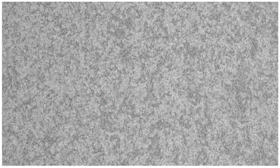
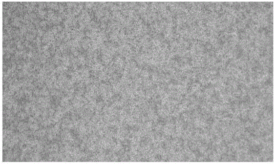
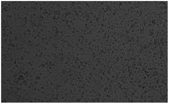
[0026] Observation of cell morphology: Most of the primary amnion epithelial cells isolated in Example 2 adhered to the wall after being cultured for 24 hours. The cells that had just adhered to the wall were elliptical and had strong light-shielding properties. Afterwards, the cytoplasm of the cells gradually expanded and the refractive properties weakened. Three days after the cells adhered to the wall, they grew rapidly, proliferated significantly, and the number of cells increased significantly, and the cells entered the exponential growth phase, such as figure 1 . A monolayer was formed in about 5 days, and the cells fused into sheets and grew in a membranous shape. The cells are flat and irregular polygonal, with clear outlines, abundant cytoplasm, and round oval cells, such as figure 2 . After subculture for about 8 hours, the cells adhere to the wall, and a good monolayer can be formed in 3 days, such as image 3 .


PUM

PropertyMeasurementUnit
Diameteraaaaaaaaaa

Abstract

The present invention discloses a culture medium for culturing human amniotic epithelial cells, wherein the culture medium comprises DMEM, fetal bovine serum, sodium pyruvate, L-glutamine, EGF, beta-mercaptoethanol and non essential amino acids. According to the present invention, the culture medium of the present invention is suitable for the culture of the human amniotic epithelial cells, especially for the culture of the primary human amniotic epithelial cells during the separation and purification processes, and the probability of immune reactions caused during the transplantation process can be reduced with the culture medium.

Description


Claims


Application Information

Patent Timeline
Owner 吉林和泽生物科技有限公司
Why Eureka
Try Eureka