Antibodies that specifically bind to TIM3

a technology of anti-tim3 and specific tim3 is applied in the field of anti-tim3 specifically binding antibodies, which can solve the problems of not effectively eliminating cancer, all members of the major categories of antineoplastic agents have considerable toxic effects on normal cells, and the inability of anti-cancer drugs to discriminate between normal and cancer cells, etc., to achieve the effect of reducing inflammation and reducing cancer cell growth

Active Publication Date: 2014-09-23
ONK THERAPEUTICS LTD
View PDF10 Cites 197 Cited by
  • Summary
  • Abstract
  • Description
  • Claims
  • Application Information

AI Technical Summary

Benefits of technology

[0008]Provided herein are TIM3-specific antibodies with improved properties, e.g., for targeting cancer cells, reducing cancer cell growth, and reducing inflammation due to CD4 T cells. In some embodiments, the invention provides an isolated antibody that specifically binds the extracellular domain of TIM3 (a TIM3-specific antibody), wherein said binding results in internalization into a TIM3-expressing cell. In some embodiments, the TIM3-specific antibody is linked to a cytotoxic agent. In some embodiments, the cytotoxic agent is selected from saporin, a taxane, a vinca alkaloid, an anthracycline, and cisplatin.
[0011]In some embodiments, the internalizing TIM3-specific antibody can bind a TIM3-expressing cell, wherein said binding inhibits proliferation of the TIM3-expressing cell. In some embodiments, the internalizing TIM3-specific antibody binds a higher percentage of peripheral blood mononuclear cells (PBMCs) from an individual having a hematopoietic cell cancer than the anti-TIM3 antibody 2E2. In some embodiments, the internalizing TIM3-specific antibody can mediate complement dependent cytotoxicity (CDC), e.g., such that more than 10, 20, 30, 40, 50% or higher percentage of TIM3 expressing cells in a sample are killed by CDC within 1 hour under appropriate conditions as described herein (e.g., with functional complement proteins, at appropriate temperature, accounting for antibody concentration relative to amount of complement and cell numbers, etc.). In some embodiments, the internalizing TIM3-specific antibody does not mediate CDC, e.g., less than 10% of TIM3 expressing cells in a sample are killed by CDC within 1 hour under appropriate conditions. In some embodiments, the internalizing TIM3-specific antibody can inhibit release of an inflammatory cytokine from an activated CD4+ T cell, compared to the release of the inflammatory cytokine in the absence of the antibody.
[0036]In some embodiments, the TIM3-specific antibody that reduces (inhibits) TIM3-expressing cell proliferation also is internalized into TIM3-expressing cells. In some embodiments, the TIM3-specific antibody that reduces TIM3-expressing cell proliferation also mediates CDC. In some embodiments, the proliferation inhibiting TIM3-specific antibody does not mediate CDC. In some embodiments, the TIM3-specific antibody that reduces TIM3-expressing cell proliferation also reduces release of an inflammatory cytokine from an activated T cell, e.g., compared to release of the inflammatory cytokine in the absence of the antibody.
[0048]The high hematopoietic cancer cell binding TIM3-specific antibodies can be used for methods of treating hematopoietic cell cancers. In some embodiments, the method of treatment can comprise administering a high hematopoietic cancer cell binding TIM3-specific antibody to an individual, thereby treating the hematopoietic cell cancer in the individual. In some embodiments, the high hematopoietic cancer cell binding TIM3-specific antibody is linked to a cytotoxic agent. In some embodiments, the high hematopoietic cancer cell binding TIM3-specific antibody is cross-linked or multimerized prior to administration. In some embodiments, the method of treatment is combined with the diagnostic method, so that individuals having a hematopoietic cell cancer that displays an epitope that is well-recognized by a particular TIM3-specific antibody, as determined in the diagnostic method, are treated with the same antibody. In this way, the cancer cells in that individual can be more effectively targeted, e.g., than in a non-targeted, or non-individualized, therapeutic method. In this context, the term “well-recognized” indicates that a high percentage of PBMC or BMMC from the individual are bound by the TIM3-specific antibody, e.g., greater than 5%, 10%, 12%, 15%, 20% or more, or that the TIM3-specific antibody binds with high intensity, e.g., higher intensity than the 2E2 antibody as determined by FACS.
[0058]Further provided are antibodies that specifically bind the extracellular domain of TIM3, wherein said binding inhibits release of an inflammatory cytokine from an activated CD4+ T cell, compared to the release of the inflammatory cytokine in the absence of the antibody. In some embodiments, the inflammatory cytokine is IL-6. In some embodiments the inflammatory cytokine is IFN-gamma. In some embodiments, the inflammatory cytokine reducing TIM3 specific antibody reduces release of the inflammatory cytokine (e.g., IL-6 or IFN-gamma) more than the 2E2 antibody.

Problems solved by technology

One of the major limitations of chemotherapy is the general inability of anticancer drugs to discriminate between normal and cancer cells.
Almost all members of the major categories of antineoplastic agents have considerable toxicity for normal cells.
However, existing cancer targets do not target CSCs.
For this reason, existing chemotherapeutic strategies, even when specifically delivered to cancer cells, do not effectively eliminate the cancer.
Risk of recurrence remains because the surviving CSCs can give rise to new cancer cells.

Method used


Image

  • Antibodies that specifically bind to TIM3
  • Antibodies that specifically bind to TIM3
  • Antibodies that specifically bind to TIM3

Examples


example 1

A. Example 1

TIM3-Specific Antibodies

[0283]Female Balb / C mice were immunized with recombinant human TIM3-Fc fusion protein four times. Two days after final injection, spleen and lymph node cells were fused with SP2 / O cells using PEG and selected in HAT medium. Hybridomas were screened by ELISA for reactivity against recombinant TIM3 protein and positive cultures were confirmed by flow cytometry for recognition of TIM3 expressing cell lines. Selected hybridoma were subcloned by limiting dilution or single cell sorting. Subclones were confirmed by flow cytometry for binding to TIM3. Heavy chain and light chain variable region sequences were amplified using reverse transcriptase polymerase chain reaction (RT-PCR) with specific primers. cDNA products were then sequenced. Table 1 (SEQ ID NOS:3-5, 8-10; 13-15, 18-20; 23-25, 28-30; 73-75, 78-80; 83-85, 88-90; 93-95, 98-100; 103-105, 108-110; 33-35, 38-40; 43-45, 48-50; 53-55, 58-60; 63-65, 68-70; 113-115, 118-120) profiles the selected anti...

example 2

B. Example 2

Binding Profile for TIM3-Specific Antibodies

[0286]The binding profile of the selected TIM3-specific antibodies was tested on various TIM3-expressing cell lines. Table 2 shows the percentage of cell binding of the indicated antibodies to Pfeiffer cells, Daudi cells, and CMK cells. TIM3 2E2 is an antibody commercially available from eBioscience®, San Diego, Calif. (see, e.g., the website and catalog available at ebioscience.com, catalog number 17-3109, Hastings et al. (2009) Eur. J. Immunol. 39:2492).

[0287]

TABLE 2Profile of Tim3-specific antibody binding to myeloid and lymphoma cell linesFACS staining (Percent binding)Cell LineDisease1.7E107.10F6.18.16C109.1G1227.2H427.12E12Tim3 2E2PfeifferNon97%92%85%14%75%97%97%Hodgkin's(Geomean =(Geomean =(Geomean =Lymphoma.439)458)308)Diffuse large91%B cellLymphomaDaudiBurkitt's95%98%67%Neg.46%92%90%Lymphoma97%CMKAcute32%NDND 2%Neg.18%19%megakaryo-cyticleukemia.AML-M7

[0288]The TIM3-specific antibodies were also tested for binding to pe...

example 3

C. Example 3

Binding of TIM3-Specific Antibodies to Cancer Patient Samples

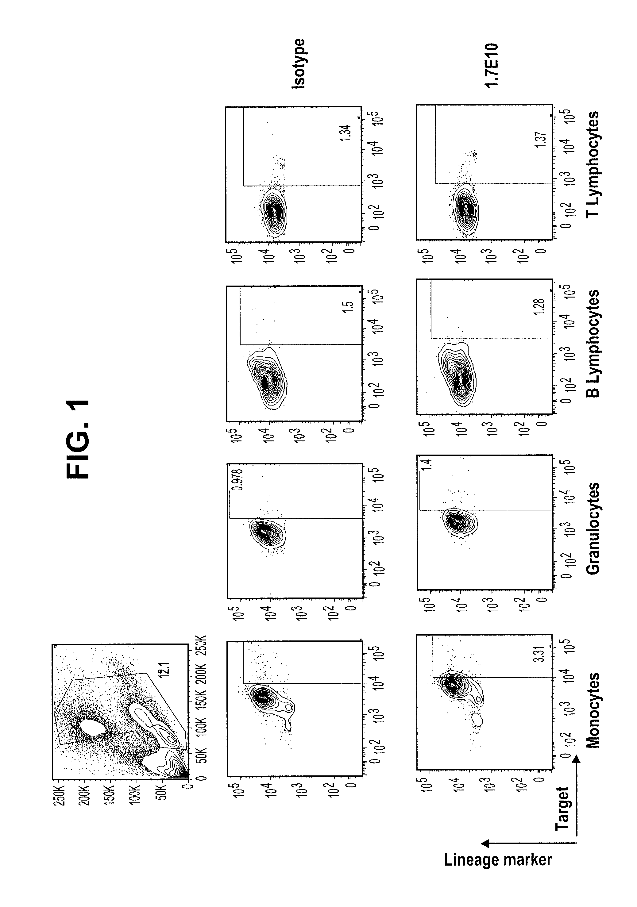
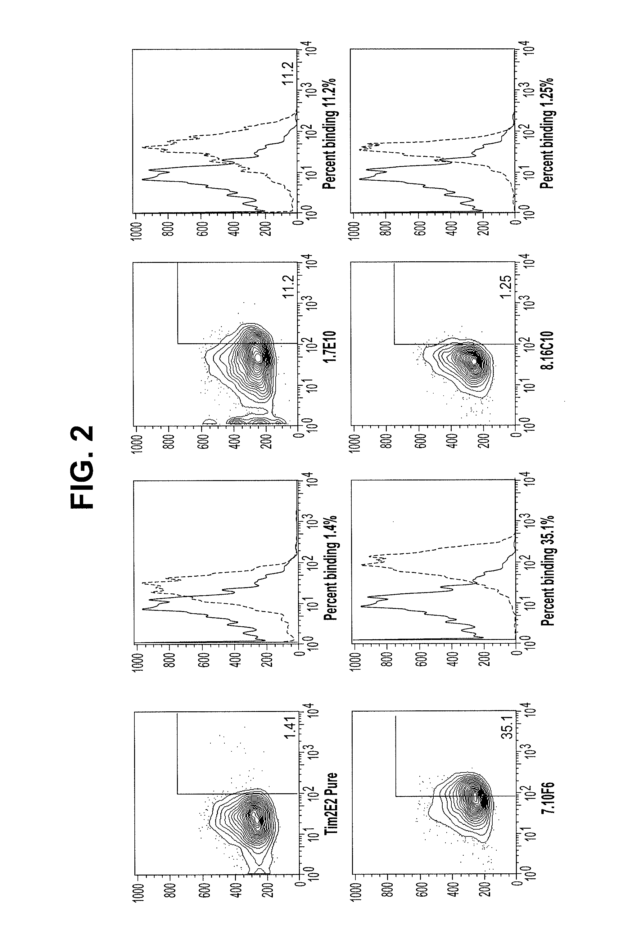
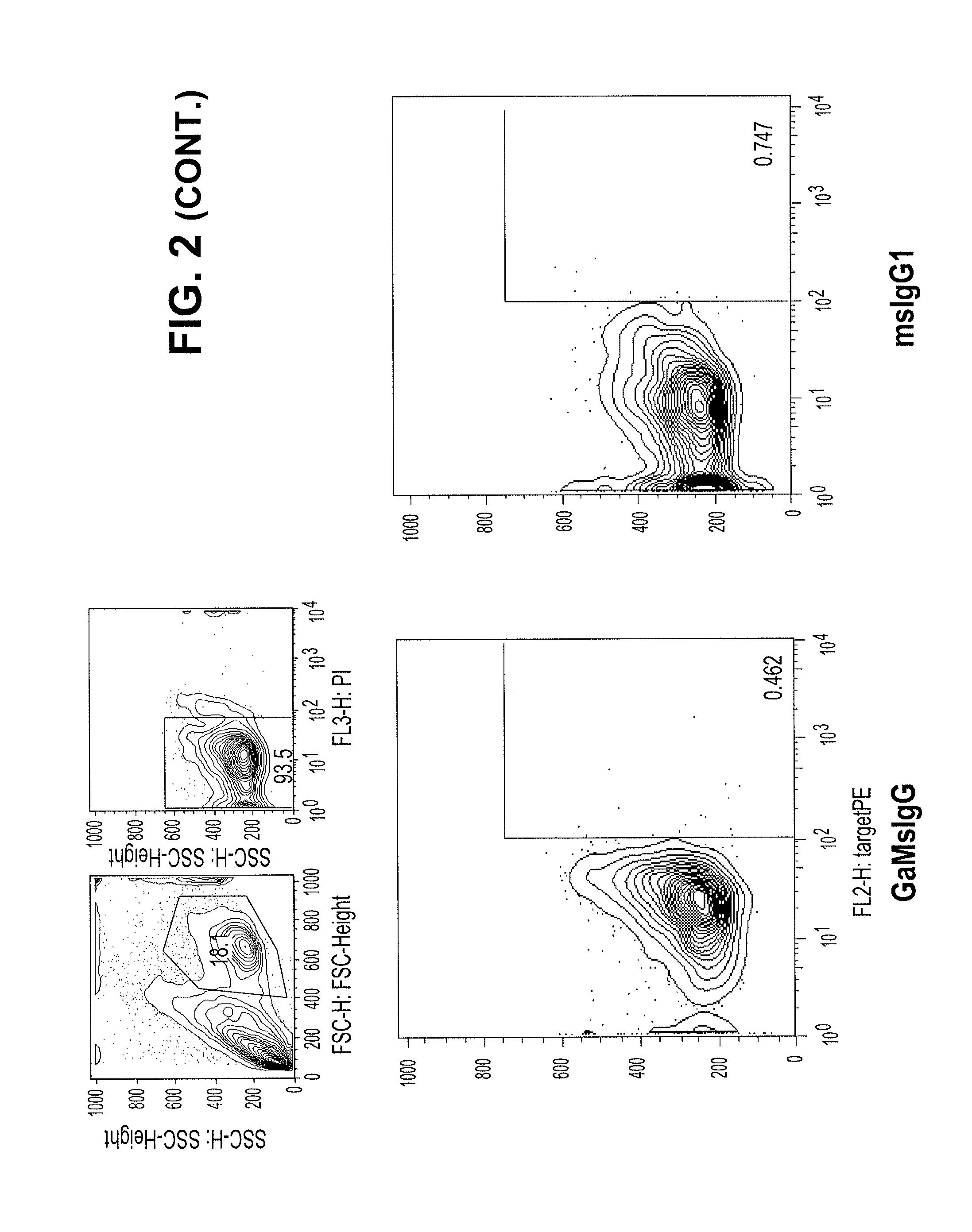
[0289]In contrast to the lack of binding to normal peripheral blood cells, the Tim3-specific antibodies bind at a high level to AML cells from patient samples. FIG. 2 compares the peripheral blood binding profiles of Isotype control (msIgG1) and 2E2 antibodies to those for the 1.7E10, 7.10F6, and 8.16C10 antibodies. The 1.7E10.7 and 7.10F6 bind to significantly more cells, with higher fluorescence, than the commercially available antibody. The 8.16C10 antibody binds about the same number of cells as 2E2 at a slightly higher fluorescence.

[0290]Table 3 shows the binding profiles for peripheral blood from five different AML patient samples. In this case, the peripheral blood was separated into stem cell (CD34+CD38−) and blast cells. Table 4 shows the binding profiles for these populations obtained from bone marrow.

[0291]

TABLE 3Percent binding of TIM3-specific antibodies to PBMCs from AML patientsIDPopulationDiagno...


PUM

PropertyMeasurementUnit
temperatureaaaaaaaaaa
concentrationaaaaaaaaaa

Abstract

Provided herein are antibodies specific for TIM3 that can be used to detect cancer cells, in particular, cancer stem cells. The antibodies can also be used in therapeutic compositions for treating cancer and reducing inflammation.

Description

CROSS-REFERENCES TO RELATED APPLICATIONS[0001]The present application claims priority to U.S. Provisional Application No. 61 / 504,062, filed Jul. 1, 2011, and U.S. Provisional Application No. 61 / 660,530, filed Jun. 15, 2012, the disclosures of which are incorporated herein by reference in their entireties.REFERENCE TO A “SEQUENCE LISTING,” A TABLE, OR A COMPUTER PROGRAM LISTING APPENDIX SUBMITTED AS AN ASCII TEXT FILE[0002]The Sequence Listing written in file -27-2.TXT, created on Aug. 28, 2012, 77,824 bytes, machine format IBM-PC, MS-Windows operating system, is hereby incorporated by reference in its entirety for all purposes.BACKGROUND OF THE INVENTION[0003]T cell immunoglobulin and mucin domain-containing molecule 3 (TIM3) is an immunoglobulin (Ig) superfamily member, expressed on Th1 cells. TIM3 has been shown to play a role in modulating the immune response of Th1 cells, and reducing inflammation in a number of conditions.[0004]TIM3 is also expressed on cancer cells, and on can...

Claims


Application Information

Patent Timeline
Patent Type & Authority Patents(United States)
IPC IPC(8): A61K39/00A61K33/243C07K16/28C07K16/30G01N33/50G01N33/574
CPCC07K16/2803G01N33/57426G01N33/57407C07K16/30C07K16/3061G01N33/5047G01N33/5073C07K2317/73C07K2317/77C07K2317/734A61K47/48561A61K31/337A61K33/24A61K38/168C07K2317/565C07K16/3092A61K31/704A61K31/4545A61K47/6849A61K31/24A61P35/00A61P35/02A61K33/243
Inventor KARSUNKY, HOLGERJIANG, YING-PING
Owner ONK THERAPEUTICS LTD
Why Eureka
Try Eureka