Conformable tissue repair implant capable of injection delivery

a tissue repair and implant technology, applied in the field of methods and apparatuses, can solve the problems of inconvenient injection delivery, inconvenient injection delivery, and high labor intensity, and achieve the effect of promoting new cellular growth and enhancing healing

Active Publication Date: 2008-01-08
DEPUY SYNTHES PROD INC
View PDF160 Cites 209 Cited by
  • Summary
  • Abstract
  • Description
  • Claims
  • Application Information

AI Technical Summary

Benefits of technology

[0009]This invention relates to a conformable tissue implant for use in treating injured or defective tissue, and a method for delivering such an implant in a minimally invasive procedure. The implant is configured to be introduced to the tissue site, where it can assume the shape or geometry of the tissue defect or injury site, thereby providing a close interface between the implant and the tissue site which enhances healing and promotes new cellular growth to occur. The biocompatible tissue implant can be used for the repair, augmentation and / or regeneration of diseased or damaged tissue. Further, the tissue implant can be used for tissue bulking, cosmetic treatments, therapeutic treatments, tissue augmentation, as well as tissue repair.
[0011]In one embodiment of the present invention, the tissue carrier matrix further includes a binding agent that acts to gel together or facilitate cohesion of the tissue fragments and granules within the tissue carrier matrix. The binding agent enables the implant to take on a semi-solid or gel-like form. Where a solid or cured implant is desired, a curing agent can additionally be provided with the tissue carrier matrix. This curing agent would act to crosslink the binding agent, thereby forming a solid implant within which are the tissue fragments and the bioresorbable, biocompatible granules. In one aspect, the implant is cured once it is delivered to the implantation site. In another aspect, the implant is cured prior to its delivery to the implantation site. To further enhance the implant's regenerative or reconstructive abilities, the tissue carrier matrix can also include a biological component or effector which enhances the effectiveness of the tissue fragments to new cellular growth.
[0013]The invention also provides a method of preparing a tissue repair implant in accordance with the present invention, which involves the steps of providing a tissue carrier matrix comprising a plurality of biocompatible, bioresorbable granules, introducing a fluid suspension containing at least one tissue fragment to the tissue carrier matrix, the tissue fragment having an effective amount of viable cells capable of migrating out of the tissue fragment and into the tissue carrier matrix, separating the at least one tissue fragment from the fluid suspension, and collecting the tissue carrier matrix with the at least one tissue fragment for implantation at a tissue site to be repaired. The tissue carrier matrix can be provided with a binding agent which enables the implant to form a gel-like or semi-solid implant. A curing agent can additionally be provided to enable the implant to set either before or after delivery to the implantation site. A biological component or an effector can also be added to the tissue carrier matrix to further enhance the effectiveness of the tissue fragments.
[0014]The tissue implant of the present invention aims to accomplish several tasks simultaneously in order to provide more efficient delivery of a tissue repair implant to a site of tissue injury or defect. The invention combines the utility of sieving or capturing a biological agent in a carrier, with the advantage of being able to immediately use the biological agent in an intraoperative procedure, in order to deliver a conformable tissue implant loaded with tissue fragments containing living cells to a tissue implantation site. Another advantage provided by the tissue repair implant of the present invention is that there is no need to isolate cells, nor is there a need to grow tissue or attach cells to the carrier prior to delivering the implant to the implantation site. Also, by using a carrier comprising bioabsorbable, biocompatible granules, the implant is able to combine sustained drug delivery capabilities and structural integrity provided by a scaffold support with the convenience of injection delivery.

Problems solved by technology

While the trend towards using tissue engineering approaches to tissue repair continues to gain popularity, mainly because of the long-term benefits provided to the patient, these current techniques are not without drawbacks.
One disadvantage with current tissue engineering techniques is that they can be time consuming.
This manual labor-intense process is extremely costly and time consuming.
Although the clinical data suggest long term benefits for the patient, the prohibitive cost of the procedure combined with the traumatic impact of two surgical procedures, has hampered adoption of this technique.
This model is limited in application because of the secondary surgery and high costs involved.
More importantly, one limitation of using such scaffolds is that tissue defect geometry can often be unpredictable.
Since the scaffold geometry is essentially limited to what has been manufactured, the scaffold carrier to be implanted rarely matches perfectly the site.
For certain difficult to match or unusually shaped sites, even the step of trimming the scaffold does not ensure an ideal fit with the implantation site.
Further, where relatively large tissue defects are involved, minimally invasive surgery may not be possible due to the limited size of the surgical access site.
Therefore, delivery of large scaffolds may require an open procedure which poses more risks to the patient.
Hence, these systems have proven to be less than ideal due to the problems associated with cell viability once incorporated into these carrier systems.

Method used


Image

  • Conformable tissue repair implant capable of injection delivery
  • Conformable tissue repair implant capable of injection delivery
  • Conformable tissue repair implant capable of injection delivery

Examples


example

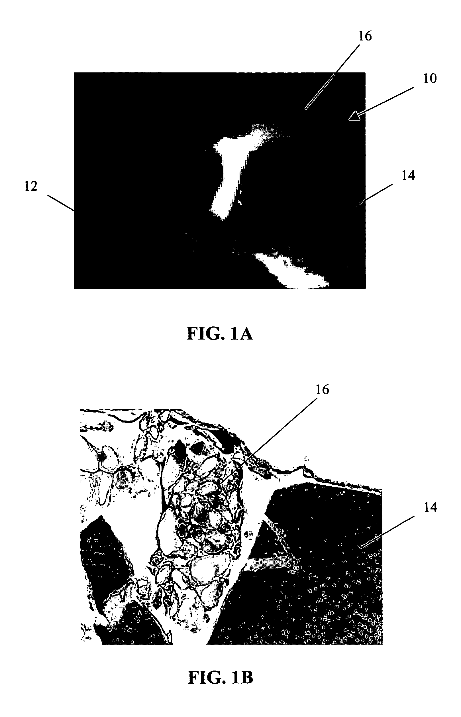
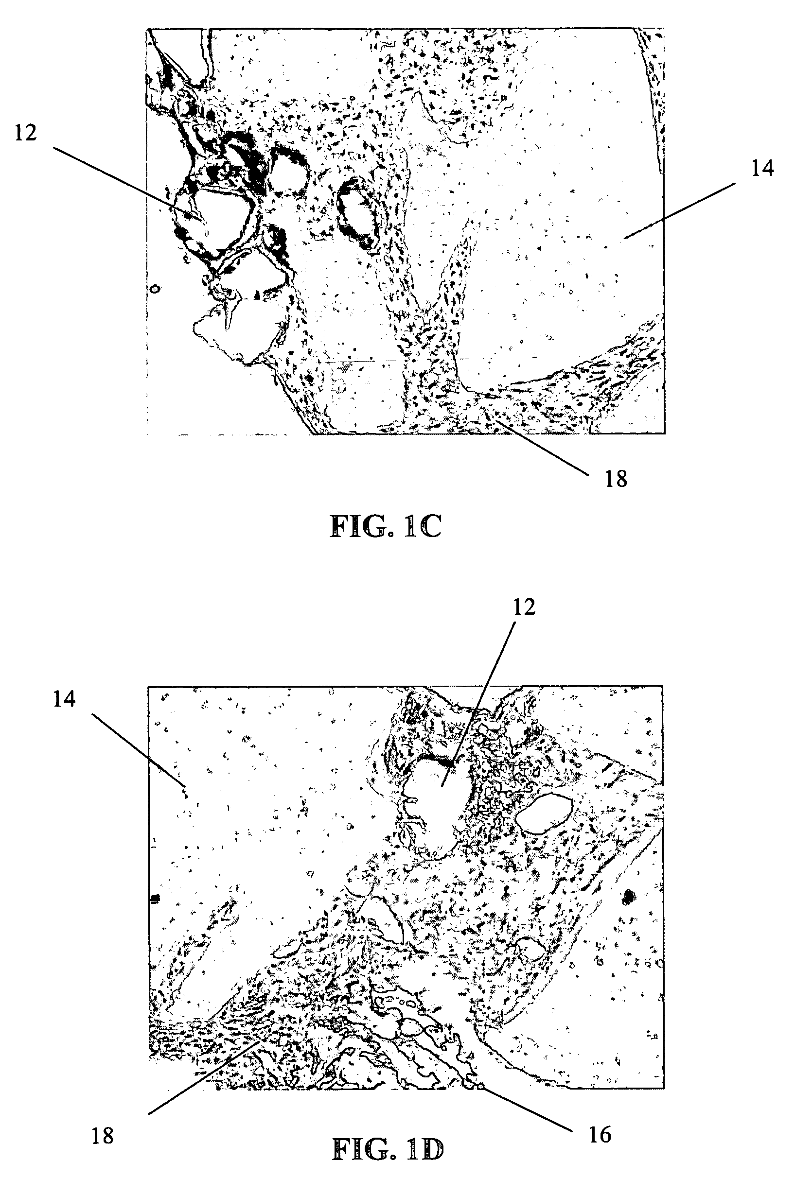
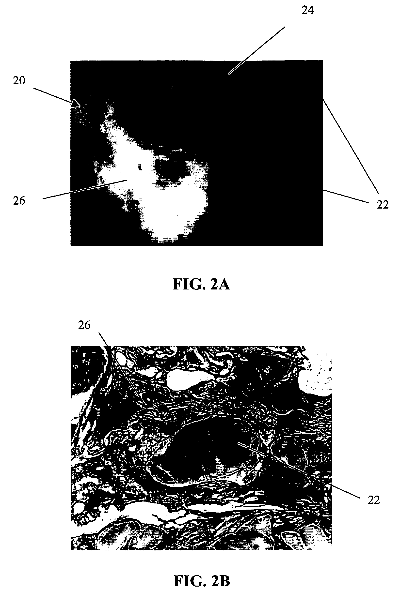
[0064]The primary objective of this study was to examine the outgrowth of chondrocytes in a sample implant comprising bovine cartilage fragments, fibrin glue and polyglycolic acid (PGA) granules in vitro. For the study, minced bovine cartilage and PGA granules of different sizes were mixed with fibrin glue (Tisseel™) and injected into a mold to form bioadhesive plugs in accordance with Table 1 below. The resulting plugs were then cultured in cell culture or chondrogenic medium for 3 and 6 week periods, respectively. After incubation, the plugs were fixed, sectioned and stained with hematoxylin and eosin (H & E).

[0065]

TABLE 1Experimental Conditions for Study of Chondrocyte Outgrowthfrom Composite Mold Containing Fibrin Glue, PLA Granules,and Bovine Cartilage FragmentsAmountIncubationof mincedAmount ofAmount ofperiodConditionscartilagePGAfibrin glue3 & 6 weeksComposite plug100 mg31 mg400 μln = 1 eachwith PGA (158 μm)granulesComposite plug100 mg31 mg400 μln = 1 eachwith PGA (286 μm)gra...


PUM

PropertyMeasurementUnit
outer diameteraaaaaaaaaa
elongationaaaaaaaaaa

Abstract

A conformable tissue implant is provided for use in repairing or augmenting a tissue defect or injury site. The tissue implant contains a tissue carrier matrix comprising a plurality of biocompatible, bioresorbable granules and at least one tissue fragment in association with the granules. The tissue fragment contains one or more viable cells that can migrate from the tissue and populate the tissue carrier matrix. Also provided is a method for injectably delivering the tissue implant.

Description

CROSS REFERENCE TO RELATED APPLICATIONS[0001]Not applicable.STATEMENT REGARDING FEDERALLY SPONSORED RESEARCH[0002]Not applicable.FIELD OF THE INVENTION[0003]The present invention relates to methods and apparatus for the treatment of tissue injuries or defects. Specifically, the present invention relates to tissue repair and augmentation implants, and more particularly, to a conformable tissue repair and augmentation implant capable of injection and a method for its minimally invasive delivery.BACKGROUND OF THE INVENTION[0004]Injuries to soft tissue, such as cartilage, skin, muscle, bone, tendon and ligament, where the tissue has been injured or traumatized frequently require surgical intervention to repair the damage and facilitate healing. Such surgical repairs can include suturing or otherwise repairing the damaged tissue with known medical devices, augmenting the damaged tissue with other tissue, using an implant, a graft or any combination of these techniques. Despite these conv...

Claims


Application Information

Patent Timeline
Patent Type & Authority Patents(United States)
IPC IPC(8): A61K35/32A61F2/02A61F2/08A61L27/00A61F2/10A61F2/28A61L27/38A61L27/58
CPCA61L27/3604A61L27/3683A61L27/38A61L27/3852A61L27/58A61P17/00A61P19/02A61P19/04A61P19/08A61P21/00A61P29/00A61P31/00A61P37/06A61P39/06A61P43/00A61P7/04
Inventor BINETTE, FRANCOISHAMMER, JOSEPH J.MUKHOPADHYAY, KRISHROSENBLATT, JOEL
Owner DEPUY SYNTHES PROD INC
Why Eureka
Try Eureka