
Large-field-of-view fundus imaging device

An imaging device and a technology with a large field of view, which can be used in ophthalmoscopes, eye testing equipment, medical science, etc., can solve problems such as difficult design and complex manufacturing processes, and achieve reduced equipment volume, reasonable device structure, and portability. The effect of functionality and practicality

Active Publication Date: 2015-03-11
TIANJIN SUOWEI ELECTRONICS TECH
View PDF12 Cites 15 Cited by
  • Summary
  • Abstract
  • Description
  • Claims
  • Application Information

AI Technical Summary

Problems solved by technology

This method is difficult to design and the manufacturing process is complicated

Method used


Image

  • Large-field-of-view fundus imaging device
  • Large-field-of-view fundus imaging device
  • Large-field-of-view fundus imaging device

Examples


Embodiment Construction

[0031] The implementation of the present invention will be described in further detail below in conjunction with the accompanying drawings. The following embodiments are only descriptive, not restrictive, and cannot limit the protection scope of the present invention.

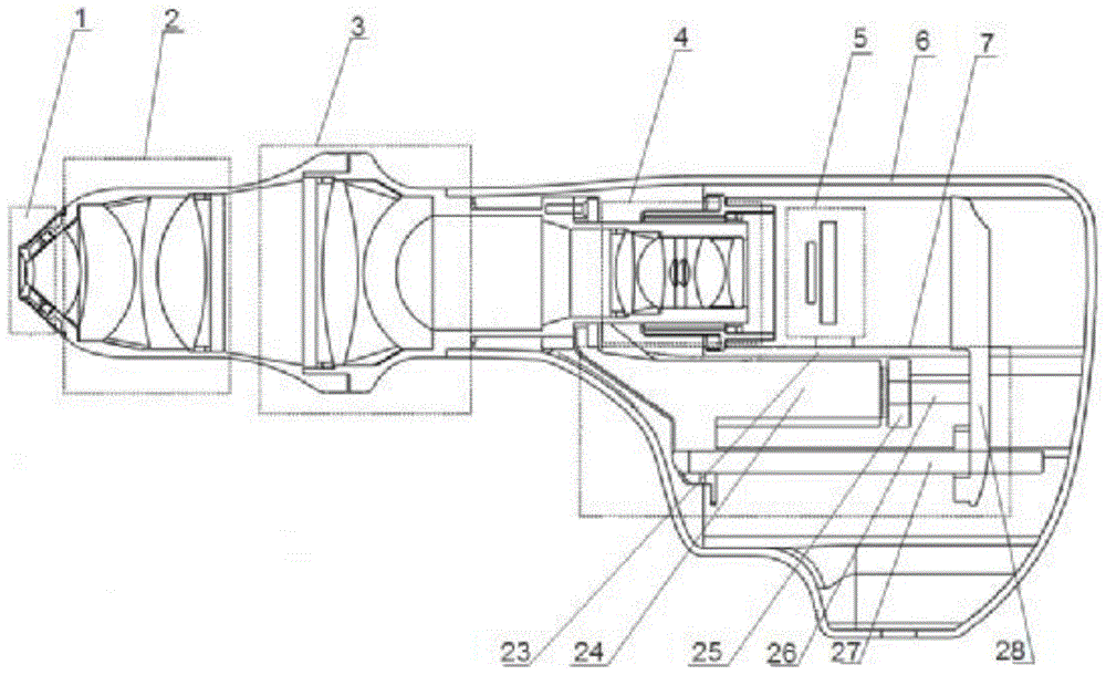
[0032] A large field of view fundus imaging device, such as figure 1 As shown, the device includes a device housing 6, and an illumination module 1, an eyepiece objective lens module 2, a field lens module 3, an imaging objective lens module 4, and an imaging As for the module 5, a focusing module 7 for adjusting the movement of the imaging module along the horizontal axis is installed below the imaging module.

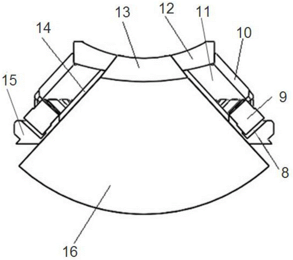
[0033] Wherein, the lighting module, such as figure 2 As shown, it includes a round platform ring-shaped light frame 15, and a plurality of center-symmetrical LED light source fixing grooves 8 are formed at regular intervals on the bottom outer side of the round platform ring-shaped light frame, and ...


PUM

PropertyMeasurementUnit
Lengthaaaaaaaaaa
Thicknessaaaaaaaaaa
Outer diameteraaaaaaaaaa

Abstract

The invention relates to a large-field-of-view fundus imaging device. The device comprises a device case, wherein a lighting module, an eye objective lens module, a field lens module, an imaging objective lens module and an imaging module are sequentially and coaxially mounted on the upper side inside the case from left to right, and a focusing module for adjusting the imaging module to move horizontally and axially is mounted below the imaging module. According to the imaging device, LEDs are used as lighting sources, the divergence angle is larger than 80 degrees, a uniform lighting field of view at 120 degrees is formed at the human eye fundus, USB3.0 data interfaces are adopted, color real-time image pickup at 20 frames per second can be performed on the fundus of a human eye after mydriasis, an image surface movement focusing mode is adopted, the overall light path structure is simplified, the device is reasonable in structure, and the size of equipment is greatly reduced.

Description

technical field [0001] The invention belongs to the technical field of ophthalmic medical equipment, and relates to a large field of view fundus imaging device using LED as an illumination source and imaging through an optical lens and CCD / CMOS. The captured images are used for the screening of fundus diseases, and are especially suitable for Early diagnosis and postoperative review of fundus retinopathy of prematurity. Background technique [0002] Retinal imaging technology has been widely used in the field of fundus detection. Medicine shows that ophthalmic diseases and systemic diseases of the body will cause lesions in the retina of the fundus in the early stage of onset. Accurate and timely detection of retinopathy can play an important role in the early diagnosis of the disease, and can also provide a good platform for the pathological research of major diseases. A complete retinal optical imaging system should be composed of fundus illumination light path and fundu...

Claims


Application Information

Patent Timeline
IPC IPC(8): A61B3/14A61B3/12
Inventor 赵培泉齐岳王熙张琨王雪乔
Owner TIANJIN SUOWEI ELECTRONICS TECH
Learn More