Automatic detection and retrieval of prior annotations relevant for imaging study for efficient viewing and reporting

An annotation and image technology, which is applied in medical reports, image generation, image analysis, etc., can solve problems such as lack, radiologists' difficulty in ROI time change/progress, and automatic detection, etc., to improve workflow efficiency. Effect

Active Publication Date: 2015-04-29
KONINKLJIJKE PHILIPS NV
View PDF5 Cites 12 Cited by
  • Summary
  • Abstract
  • Description
  • Claims
  • Application Information

AI Technical Summary

Problems solved by technology

Also, current radiology image viewing systems cannot automatically detect previous correlation studies related to the current ROI
Radiologists have difficulty accurately determining ROI change / progression over time due to lack of image linkage on a specific basis

Method used


Image

  • Automatic detection and retrieval of prior annotations relevant for imaging study for efficient viewing and reporting
  • Automatic detection and retrieval of prior annotations relevant for imaging study for efficient viewing and reporting
  • Automatic detection and retrieval of prior annotations relevant for imaging study for efficient viewing and reporting

Examples


Embodiment Construction

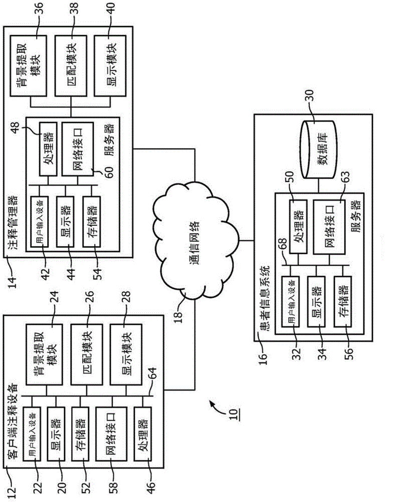
[0017] During medical imaging, one or more images are generated from a scan of a patient. Types of medical images include magnetic resonance (MR or MRI), computed tomography (CT or CAT), X-rays, ultrasound, positron emission tomography (PET) images, single photon emission computed tomography (SPECT), and others. After scans have been completed, it is common practice for radiologists to annotate the images with information that provides descriptive or identifying information for the images. The annotations provide background about the image, statistical information about the image (such as identification of the patient, type of exam, hospital, date of exam, type of acquisition, type of scan, orientation of the image, use of specific image processing filters, and Statistics associated with the region of interest) and other information. Typically, annotations include textual information, however, it should be appreciated that the annotations include various visual information, i...


PUM


Abstract

An annotation support system (10) comprising at least one display device (20). A context extraction module (24, 36) determines a context of a current medical image from a current image study. A matching module (26, 38) compares the context of the current medical image to contexts of prior medical images from prior image studies. A display module (28, 40) displays at least one of context relevant annotations and context relevant medical images from the prior image studies which match the context of the current medical study.

Description

technical field [0001] The present application generally relates to a system and method for annotating and / or retrieving content. This application is particularly concerned with, and will be described with specific reference to, the efficient retrieval of previous image studies relevant to the present study. However, it should be understood that the present application is also applicable to other usage scenarios and is not necessarily limited to the above-mentioned applications. Background technique [0002] On a routine basis, radiologists process an increasing number of images to optimally diagnose and treat patients. Patients (eg, cancer patients) undergo imaging exams frequently and accumulate many imaging studies, often involving the same anatomical region, in their medical records over time. When a radiologist reads a current exam, he or she compares the current exam images with images from previous exams in order to determine how lesions have changed over time and t...

Claims


Application Information

Patent Timeline
Patent Type & Authority Applications(China)
IPC IPC(8): G06F19/00G16H10/60
CPCG16H50/70G16H15/00G16H30/40G06T11/60G06T7/00G06T2210/41G16H30/20
Inventor T·D·D·S·马博杜瓦纳钱悦晨M·塞芬斯特G·R·曼科维奇
Owner KONINKLJIJKE PHILIPS NV
Why Eureka
Try Eureka