A Tissue Characterization Method Based on Ultrasound Radio Frequency Time Series

A time-series and ultrasound technology, applied in the fields of ultrasound/sonic/infrasonic Permian technology, ultrasound/sonic/infrasonic image/data processing, organ movement/change detection, etc. , There are differences in tissue attenuation ability, etc.

Active Publication Date: 2018-06-29
SOUTH CHINA UNIV OF TECH
View PDF3 Cites 0 Cited by
  • Summary
  • Abstract
  • Description
  • Claims
  • Application Information

AI Technical Summary

Problems solved by technology

The ultrasonic B-mode image characterization method has the following defects: (1) the brightness modulation is used to display the amplitude information, and the rich phase information is discarded, so that part of the tissue information is lost; (2) the method uses the gray scale of the ultrasonic image , the inspection results are affected by the instrument model, instrument settings, etc., and the consistency is poor
This method requires deep attenuation compensation, and the tissue attenuation capabilities of different lesions are different. At the same time, individual differences will also make the sound wave propagation path different, which will seriously affect its classification accuracy.
[0004] The above-mentioned ultrasonic tissue characterization methods are sensitive to noise and are greatly affected by individual differences. The extracted features are often only texture features or spectral features. The angle of feature extraction is too single and the amount of information is not rich enough.

Method used


Image

  • A Tissue Characterization Method Based on Ultrasound Radio Frequency Time Series
  • A Tissue Characterization Method Based on Ultrasound Radio Frequency Time Series
  • A Tissue Characterization Method Based on Ultrasound Radio Frequency Time Series

Examples


Embodiment

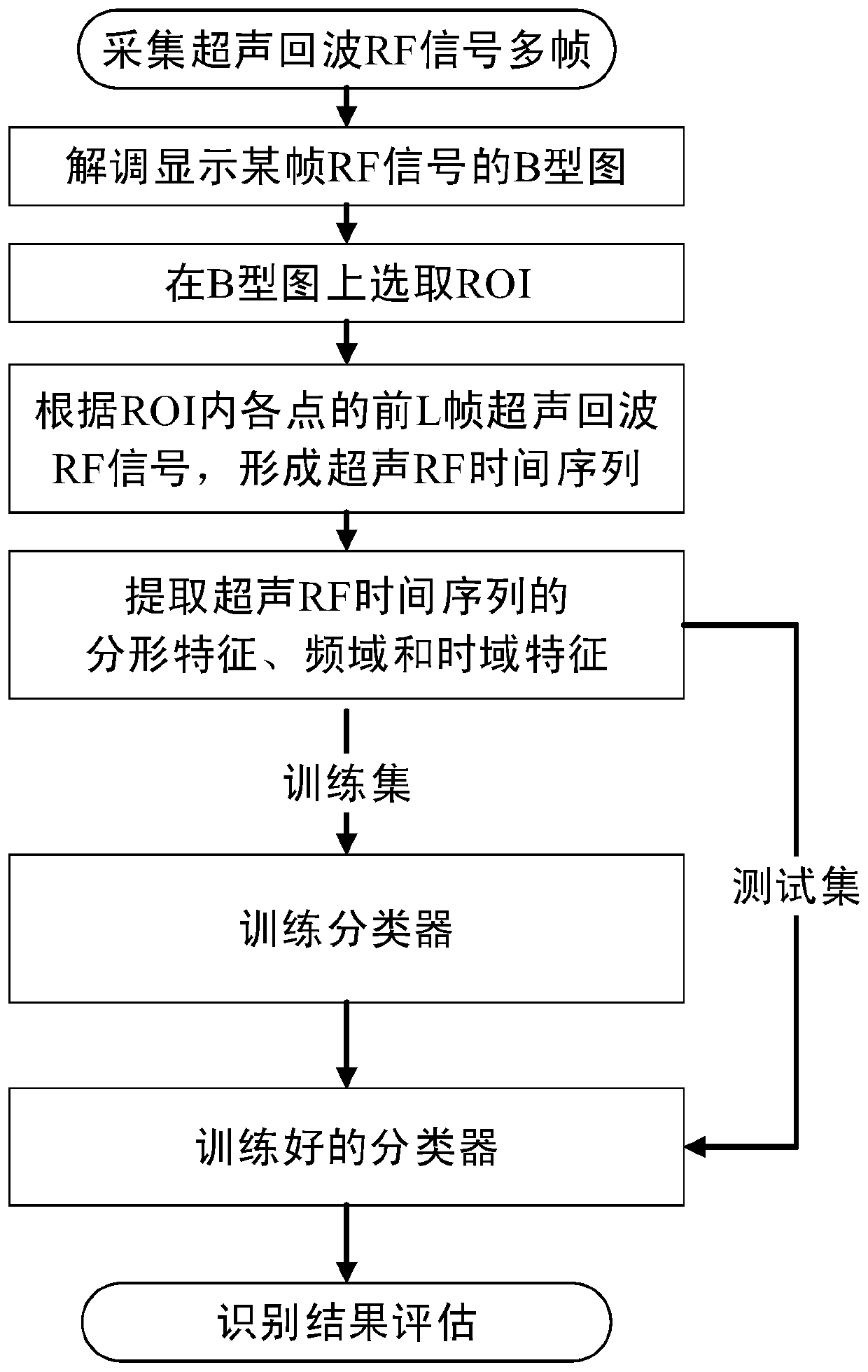
[0086] Such as figure 1 As shown, in this embodiment, a tissue characterization method based on ultrasonic radio frequency time series comprises the following steps:

[0087] S1. Construction of ultrasonic RF time series.

[0088] S1.1 Use Sonix TOUCH produced by Canada Ultrasonix Company and a broadband linear array ultrasound probe with a center frequency of 6.6MHz to scan the liver tissue area under the liver capsule of Wistar rats, and record multiple frames of ultrasound echo RF signals.

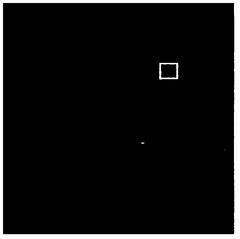
[0089] S1.2 demodulates the data of the 100th frame and displays its ultrasonic B-type image, such as figure 2 shown.

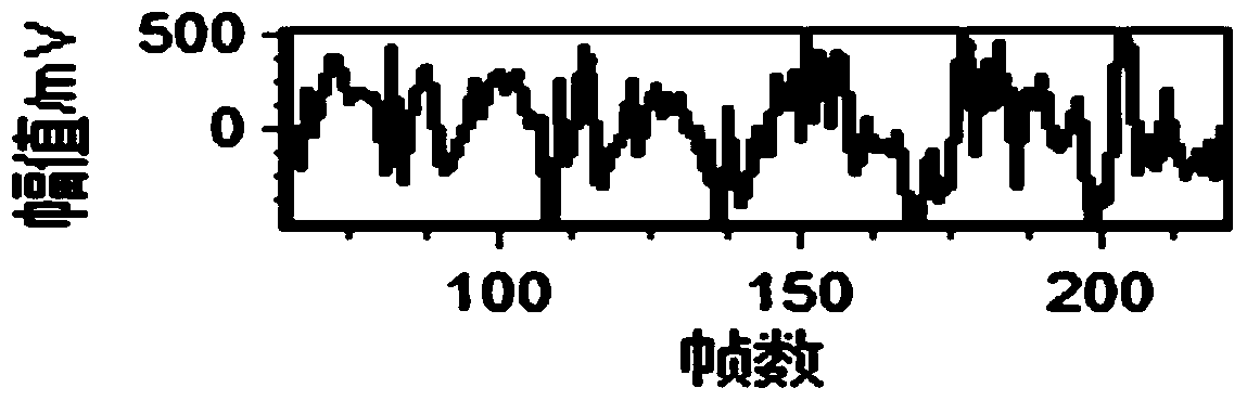
[0090] S1.3 Select a ROI with a size of 70×20 on the ultrasonic B-type map, intercept and obtain ultrasonic echo RF signals of 1400 points in the ROI, and take the first 256 frames of data for each point in the ROI to obtain 1400 points. 256 ultrasound RF time series, the ultrasound RF time series diagram is as follows image 3 shown.

[0091] S2. Feature extract...


PUM


Abstract

The invention discloses an ultrasonic RF (radio frequency) time sequence-based tissue characterization method, which comprises the following steps of 1, feature extraction: performing operations as shown as follows on each tissue sample to obtain corresponding feature vectors; 2, tissue recognition, wherein the step1 comprises the following steps: 1-1, scanning tissues to acquire multiple frames of ultrasonic echo RF signals of the tissues; 1-2, demodulating a certain ultrasonic echo RF signal and displaying a B-mode graph; 1-3, selecting an ROI (region of interest) of which the size is M*N from the B-mode graph; 1-4, taking previous L RF signals from each point in the ROI to construct M*N ultrasonic RF time sequences of which the length is L; 1-5, extracting fractal features, frequency domain features and time domain features of the ultrasonic RF time sequences to obtain the feature vectors of each tissue samples. According to the ultrasonic RF time sequence-based tissue characterization method disclosed by the invention, the features from three angles: fractal, frequency domain and time domain, are extracted based on the ultrasonic RF time sequences, and the tissues are recognized by using a classifier; the ultrasonic RF time sequence-based tissue characterization method can be widely applied to the field of ultrasonic medical detection and ultrasonic tissue characterization, and the like.

Description

technical field [0001] The invention relates to the fields of ultrasonic medicine and ultrasonic tissue characterization, in particular to a tissue characterization method based on ultrasonic radio frequency time series. Background technique [0002] Due to the complexity of the transmission and reflection characteristics of ultrasound through tissue, the interaction mechanism between ultrasound and tissue is not yet fully understood. People can only indirectly achieve the purpose of distinguishing normal and diseased tissue by obtaining useful information about tissue and explaining it. Other diagnostic methods are invasive, irreproducible, and prone to complications, which prompt people to conduct research on ultrasonic tissue characterization. [0003] There are mainly two traditional ultrasonic tissue characterization methods: ultrasonic B-mode image and ultrasonic echo RF signal. Tissue characterization based on ultrasound B-mode images mainly extracts the texture feat...

Claims


Application Information

Patent Timeline
Patent Type & Authority Patents(China)
IPC IPC(8): A61B8/08
Inventor 高永振林春漪
Owner SOUTH CHINA UNIV OF TECH
Why Eureka
Try Eureka