Breast cancer pathology image mitosis nucleus automatic segmentation method

A mitotic and automatic segmentation technology, applied in the field of image processing, which can solve the problems of unbalanced distribution of positive and negative samples, difficulty and so on

Active Publication Date: 2016-06-29
湖南品信生物工程有限公司
View PDF3 Cites 15 Cited by
  • Summary
  • Abstract
  • Description
  • Claims
  • Application Information

AI Technical Summary

Problems solved by technology

A large number of image segmentation methods such as threshold segmentation, watershed segmentation, morphological methods, and LoG-based blob detection have been used to segment cell nuclei. However, these algorithms are too simple, and often segment a large number of mi...

Method used


Image

  • Breast cancer pathology image mitosis nucleus automatic segmentation method
  • Breast cancer pathology image mitosis nucleus automatic segmentation method
  • Breast cancer pathology image mitosis nucleus automatic segmentation method

Examples


Embodiment Construction

[0056] The present invention will be further described below in conjunction with the accompanying drawings and embodiments.

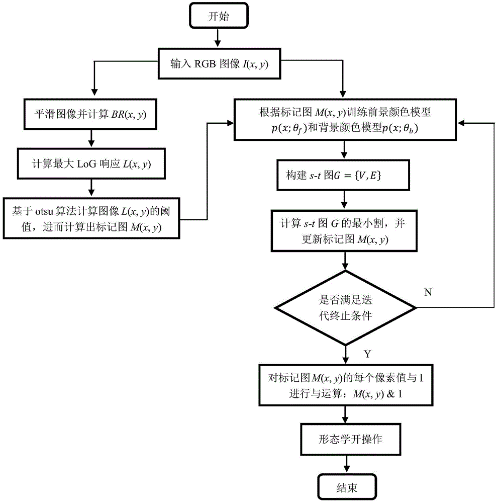
[0057] Such as figure 1 As shown, the method of the present invention mainly includes the following steps: preprocessing the original image I (x, y), and calculating its BR image; filtering the BR image with a group of LoG filters of different scales, and calculating the maximum response image L (x, y); Calculate the global threshold of the maximum response image according to the otsu algorithm, and then calculate the marker map M(x, y); collect the pixels on the image I(x, y) according to the marker map M(x, y), respectively Train the foreground Gaussian mixture color model p(x; θ f ) and background Gaussian mixture color model p(x; θ b ); construct the s-t graph G={V,E} according to the image I(x, y), the marker graph M(x, y) and the Gaussian mixture model; calculate the maximum flow of the s-t graph G to segment the graph G, and update the marker ...


PUM


Abstract

The invention discloses a breast cancer pathology image mitosis nucleus automatic segmentation method. A foreground pixel and a background pixel can be roughly estimated according to a maximum response of LoG filters of different scales of a BR graph. The color distribution of the foreground pixel and the background pixel can be described by Gaussian Mixed Models GMM, which are trained respectively according to the current foreground pixel and the current background pixel. An s-t graph G=(V,E) can be established, and the new estimation of the foreground and the background can be realized by calculating the maximum flow of the graph G, and then the GMM training and the Graph-Cut segmentation can be repeated, till the convergence is realized, or the predetermined iterations can be satisfied. The morphology open operation of the segmentation result can be carried out, and the final segmentation can be completed. The breast cancer pathology image mitosis nucleus automatic segmentation method is advantageous in that during the segmentation, the probability of the single pixel color characteristic belonging to the foreground or the background can be considered, and the relation between the adjacent pixels can be considered, and therefore the smoothness of the segmentation results can be guaranteed, and the precision can be improved.

Description

technical field [0001] The invention belongs to the technical field of image processing, in particular to a method for automatically segmenting mitotic nuclei in pathological images of breast cancer. Background technique [0002] According to statistics from the Ministry of Health, breast cancer accounts for a large proportion of cancers in women in my country, and has become one of the biggest killers that endanger women's lives. The age of onset ranges from 20 to 70 or 80 years old, and the incidence It tends to increase with age. At present, most breast cancer patients in our country are concentrated in the age group of 45-55, and the incidence rate accounts for 7-10% of all kinds of malignant tumors in the whole body, which is second only to the incidence rate of uterine cancer in women. At present, the etiology of breast cancer is not completely clear, and its early detection and early diagnosis are very important. [0003] Mitotic count is an important indicator in th...

Claims


Application Information

Patent Timeline
IPC IPC(8): G06T7/00
CPCG06T2207/20081G06T2207/30096
Inventor 梁毅雄陈再良廖胜辉王磊向遥郭璠邹北骥
Owner 湖南品信生物工程有限公司
Why Eureka
Try Eureka