
Method for positioning organs in medical image

A medical image and image technology, applied in the field of image processing in the medical field, can solve the problems of high computational complexity and complex work in recognition and positioning

Active Publication Date: 2016-07-06
SHANGHAI UNITED IMAGING HEALTHCARE
View PDF4 Cites 12 Cited by
  • Summary
  • Abstract
  • Description
  • Claims
  • Application Information

AI Technical Summary

Problems solved by technology

[0002] Existing methods for identifying and locating organs and tissues in 3D CT images, such as methods based on machine learning, require complex preliminary work, involving a large number of training image collection and preprocessing, and require the design of complex classifiers, and the computational complexity of identification and locating higher

Method used


Image

  • Method for positioning organs in medical image
  • Method for positioning organs in medical image
  • Method for positioning organs in medical image

Examples


Embodiment Construction

[0030] The present invention will be described in further detail below in conjunction with the accompanying drawings and specific embodiments. Advantages and features of the present invention will be apparent from the following description and claims. It should be noted that the drawings are all in a very simplified form and use imprecise ratios, which are only used to facilitate and clearly assist the purpose of illustrating the embodiments of the present invention.

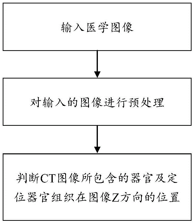
[0031] Please refer to figure 1 As shown, the method for locating organs on medical images in the embodiment of the present invention is a method for locating organs on CT images, which specifically includes the following steps:

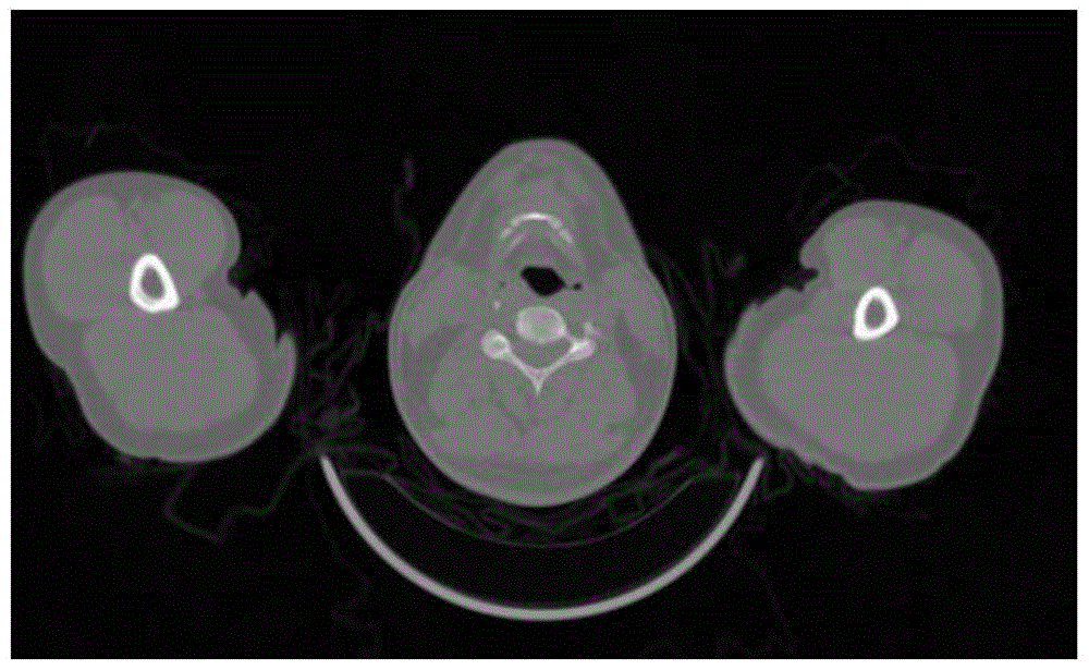
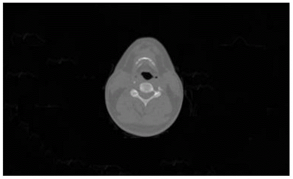
[0032] Step S1: Input a medical image including several slice images arranged along the Z direction, the medical image may be a CT image or a magnetic resonance imaging image, and the CT image contains volume data. The Z direction may be a head-to-toe direction.

[0033] Step S2: Pr...


PUM


Abstract

The invention discloses a method for positioning organs in a medical image. The method comprises following steps of step S1, inputting the medical image containing a plurality of slice images; step S2, preprocessing the input images; filtering non-body pixels; step S3, calculating the number of connected domains of each layer of slice image; through judging the numbers and the positions of the connected domains, removing the parts except first organs; confirming that the medical image contains the first organs; calculating the feature value of each layer of slice image, thus obtaining a plurality of feature values; forming a feature curve by the plurality of feature values and the slice number of the slice images; wherein the step of calculating the feature value comprises with respect to each layer of slice image, calculating the proportion of a pixel area of the pixel gray value or CT value relative to the whole body pixel area in a first range; and positioning the slice images in which the organs are located according to the proportions. According to the settings, the ranges in which the organs are located can be positioned rapidly, and the accuracy is high.

Description

technical field [0001] The invention relates to image processing in the medical field, in particular to a method for locating organs and tissues in a three-dimensional CT image. Background technique [0002] Existing methods for identifying and locating organs and tissues in 3D CT images, for example, methods based on machine learning, this method involves complex preliminary work, involves a large number of training image collection and preprocessing, and requires the design of complex classifiers, and the computational complexity of identification and locating higher. In many image processing applications, such as segmentation, registration, automatic recognition of image parts, rough image alignment, etc., it is necessary to perform preliminary identification and judgment on image parts or organs and tissues. Easy way to do it. [0003] Therefore, it is necessary to improve the existing methods for locating organs and tissues in three-dimensional CT images, and to impro...

Claims


Application Information

Patent Timeline
IPC IPC(8): G06T7/00G06T7/11G06T7/187
CPCG06T2207/10081G06T2207/10088G06T2207/30012G06T2207/30016G06T2207/30061
Inventor 黎维娟马杰延
Owner SHANGHAI UNITED IMAGING HEALTHCARE
Learn More