Bone segmentation from image data

An image data set and data technology, applied to the specific application of computer tomography to describe, the field of bone segmentation, can solve the problem of increasing excessive noise

Active Publication Date: 2016-07-20
KONINKLJIJKE PHILIPS NV
View PDF8 Cites 6 Cited by
  • Summary
  • Abstract
  • Description
  • Claims
  • Application Information

AI Technical Summary

Problems solved by technology

A potential challenge is that some skeletal structures have very similar attenuation and spectral properties to other structures in the body (e.g. contrast-enhancing organs)
In addition, spectral CT research suffers from the inherent excessive noise problem that further amplifies the challenge

Method used


Image

  • Bone segmentation from image data
  • Bone segmentation from image data
  • Bone segmentation from image data

Examples


Embodiment Construction

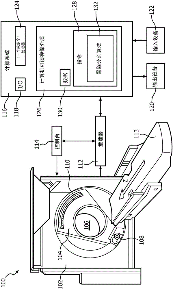
[0018] figure 1 An imaging system 100 is shown schematically, such as a computed tomography (CT) scanner. Imaging system 100 includes a stationary gantry 102 and a rotating gantry 104 . The rotating gantry 104 is rotatably supported by the stationary gantry 102 and rotates about a longitudinal or z-axis (“Z”) about an inspection zone 106 .

[0019] A radiation source 108 (eg, an x-ray tube) is rotatably supported by and rotates with the rotating gantry 104 and emits multi-energy radiation throughout the examination region 106 . In the illustrated embodiment, radiation source 108 comprises a single broad spectrum x-ray tube. In a variation, the radiation source 108 is configured to controllably switch between at least two different emission voltages (eg, 80 kVp, 140 kVp, etc.) during a scan. In another variation, radiation source 108 includes two or more x-ray tubes configured to emit radiation having different mean spectra. In another variation, the radiation source 108 in...


PUM


Abstract

A method for segmenting bone in spectral image data is described herein. The spectral image data includes at least a first set of image data corresponding to a first energy and second set of image data corresponding to a second different energy. The method includes obtaining the spectral image data. The method further includes extracting a set of features for each voxel in spectral image data. The method further includes determining, for each voxel, a probability that each voxel represents bone structure based on the set of features. The method further includes extracting bone structure from the spectral image data based on the probabilities.

Description

technical field [0001] The following generally relates to image data processing, and more specifically to bone segmentation based on image data, and is described in conjunction with a specific application of computed tomography (CT). However, the following may also apply to other imaging modalities. Background technique [0002] A CT scanner typically includes an x-ray tube mounted on a rotatable gantry positioned across the examination region opposite the detector array. The rotatable gantry and x-ray tube rotate around the examination area. The x-ray tube emits radiation that traverses the examination region and is detected by the detector array. The detector array generates and outputs signals indicative of detected radiation. The signal is reconstructed to generate three-dimensional volumetric image data. The volumetric image data includes voxels represented by grayscale intensity values ​​corresponding to relative radiance intensities. [0003] The grayscale values...

Claims


Application Information

Patent Timeline
IPC IPC(8): G06T7/00
CPCG06T2207/10081G06T2207/20076G06T2207/30008G06T7/11G06T7/143G16H30/20G06T5/70
Inventor L·戈申A·格林高兹
Owner KONINKLJIJKE PHILIPS NV
Why Eureka
Try Eureka