Single-port thoracoscope incision separation fixator

A fixator and thoracoscopic technology, applied in the field of medical devices, can solve problems such as long duration, aggravated patient discomfort, and poor accuracy, and achieve the effects of reducing fatigue, avoiding implant transfer, and avoiding mutual interference

Inactive Publication Date: 2016-12-21
中国医科大学附属第四医院
View PDF5 Cites 3 Cited by
  • Summary
  • Abstract
  • Description
  • Claims
  • Application Information

AI Technical Summary

Problems solved by technology

[0003] Single-port thoracoscopic surgery itself also has certain disadvantages. The existing thoracic-related equipment is not suitable for single-port thoracoscopic operation: one operation hole, all operating instruments enter and exit through this port, there is mutual interference between instruments, and there is often a After the instrument is inserted, other instruments cannot enter or cannot move; it is difficult for the hand holding the mirror to fix the light source of the thoracoscope, the accuracy is poor, and it is easy to fatigue; the smoke generated by the electric knife or electrocoagulation cannot be discharged smoothly (because it cannot be put into In addition, thoracoscopic surgical instruments are relatively hard and have a large diameter, and they repeatedly enter and exit from the intercostal space during the operation, which aggravates the damage to the intercostal nerves and causes postoperative discomfort for patients aggravated and prolonged
[0004] For the technical problems related to single-port thoracoscopic surgery, no effective solutions have been proposed so far

Method used


Image

  • Single-port thoracoscope incision separation fixator
  • Single-port thoracoscope incision separation fixator
  • Single-port thoracoscope incision separation fixator

Examples


Embodiment Construction

[0015] The following will clearly and completely describe the technical solutions in the embodiments of the present invention with reference to the accompanying drawings in the embodiments of the present invention. Obviously, the described embodiments are only some, not all, embodiments of the present invention. All other embodiments obtained by persons of ordinary skill in the art based on the embodiments of the present invention belong to the protection scope of the present invention.

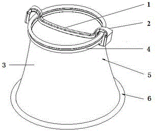
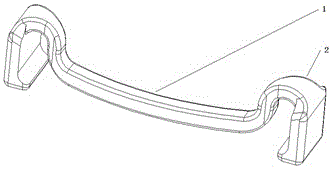
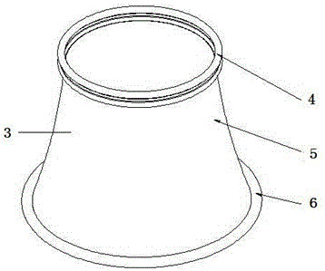
[0016] Such as Figure 1-3 As shown, a single-hole thoracoscopic incision partition fixer according to an embodiment of the present invention includes an incision retractor fixer 3 and an incision partition fixation clip 1 . The incision retracting fixture 3 is put into the chest incision, and the incision is retracted due to the elasticity of the retracting fixture 3, and the incision is separated and fixed to the outside of the clip 1 through the buckle 2 and the incision retracting fixture...


PUM


Abstract

The invention discloses a single-port thoracoscope incision separation fixator which comprises an incision retraction fixator and an incision separation fixing clamp retainer. The incision separation fixing clamp retainer is connected with an outer clamp ring of the incision retraction fixator through a buckle. The incision retraction fixator comprises an inner elastic clamp ring, an outer elastic clamp ring and a connecting pipeline which are integrated. The incision separation fixing clamp retainer is connected with the outer clamp ring of the incision retraction fixator to divide the space of an incision into two operation ducts. The single-port thoracoscope incision separation fixator has the advantages that by arranging the incision retraction fixator and the incision separation fixing clamp retainer, the single-port thoracoscope incision separation fixator has the effects of a single-port thoracoscope surgery operation platform; furthermore, the single-port thoracoscope incision separation fixator is convenient to use and practical, and single-port thoracoscope operation can be achieved.

Description

technical field [0001] The invention relates to the technical field of medical instruments, more precisely, it relates to a single-hole thoracoscopic incision dividing fixator. Background technique [0002] Minimally invasive surgery is the direction of surgical development, and its purpose is to reduce the pain of surgery and improve the quality of life of patients. The traditional 3-incision video-assisted thoracoscopic surgery (VATS) has reached a consensus on the therapeutic effect of thoracic diseases, and has become an essential technique for thoracic surgeons. The exploration of "minimally invasive" thoracic surgery in single-port thoracoscopic surgery is a new direction of minimally invasive thoracic surgery. Single-port thoracoscopic surgery refers to the use of only one incision of about 2.0-4.0 cm for thoracoscopic surgery, and the observation mirror and surgical instruments pass through the same surgical incision. The clinical effect of single-port thoracoscopi...

Claims


Application Information

Patent Timeline
Patent Type & Authority Applications(China)
IPC IPC(8): A61B17/00A61B17/02
CPCA61B17/00234A61B17/0218A61B2017/00238A61B2017/00309A61B2017/00336A61B2017/0034
Inventor 杨雪鹰黄焱
Owner 中国医科大学附属第四医院
Why Eureka
Try Eureka