
Fundus image classification method and device of retinopathy

A technology for retinopathy and fundus images, applied in the field of image processing, can solve the problems of high requirements for doctors' medical resources and heavy workload of doctors, and achieve the effect of reducing high standard requirements, reducing workload and improving accuracy

Inactive Publication Date: 2017-03-22
CAPITAL UNIVERSITY OF MEDICAL SCIENCES
View PDF6 Cites 73 Cited by
  • Summary
  • Abstract
  • Description
  • Claims
  • Application Information

AI Technical Summary

Problems solved by technology

[0004] However, in the above-mentioned diagnostic method, it is necessary to manually classify and process a large number of fundus photo images, which makes the doctor's workload larger and the above-mentioned diagnostic method requires a higher level of medical resources for the doctor.

Method used


Image

  • Fundus image classification method and device of retinopathy
  • Fundus image classification method and device of retinopathy
  • Fundus image classification method and device of retinopathy

Examples


Embodiment Construction

[0057] In order to make the purposes, technical solutions and advantages of the embodiments of the present invention clearer, the technical solutions in the embodiments of the present invention will be clearly and completely described below with reference to the drawings in the embodiments of the present invention. Obviously, the described embodiments are only These are some embodiments of the present invention, but not all embodiments. The components of the embodiments of the invention generally described and illustrated in the drawings herein may be arranged and designed in a variety of different configurations. Thus, the following detailed description of the embodiments of the invention provided in the accompanying drawings is not intended to limit the scope of the invention as claimed, but is merely representative of selected embodiments of the invention. Based on the embodiments of the present invention, all other embodiments obtained by those skilled in the art without c...


PUM


Abstract

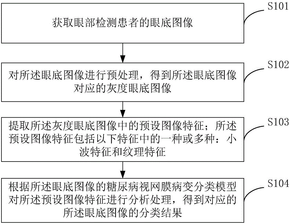
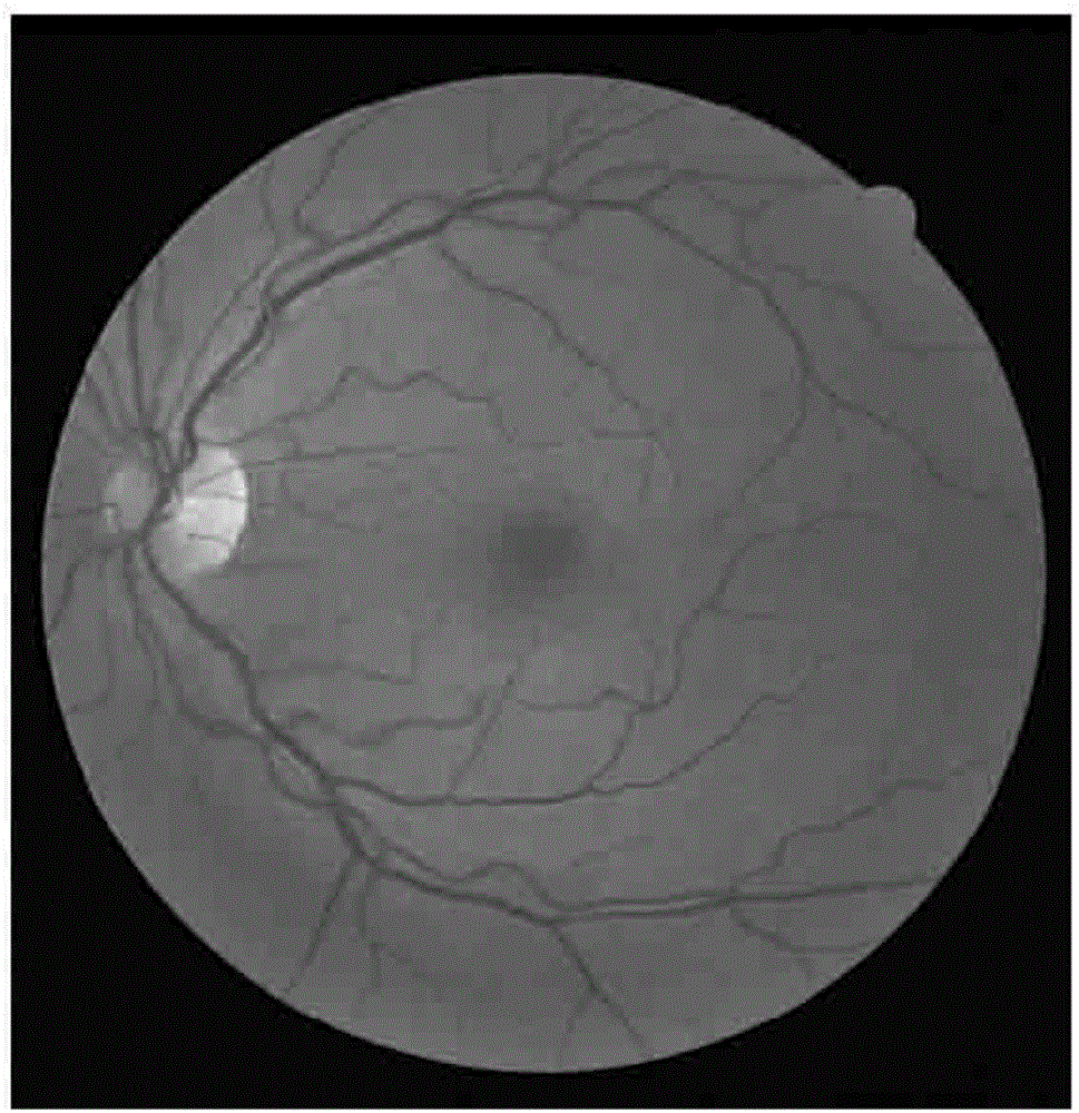
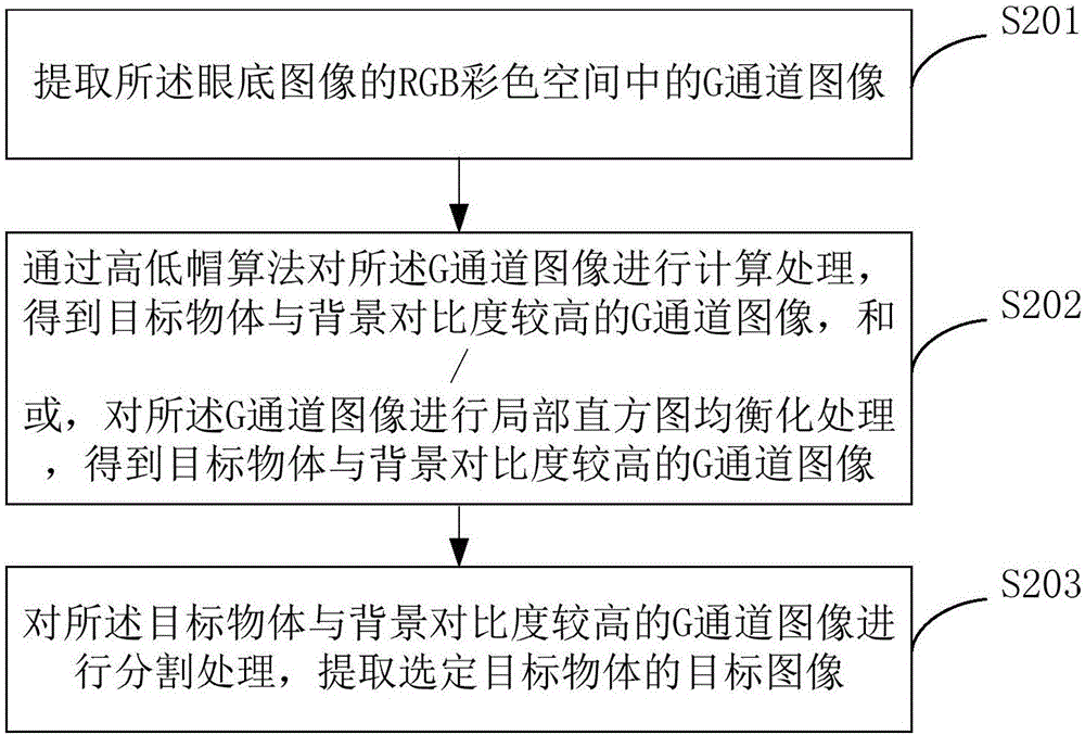
The present invention provides a fundus image classification method and device of retinopathy. The method comprises a step of obtaining the fundus image of an eye detection patient, a step of preprocessing the fundus image and obtaining a corresponding grey fundus image, a step of extracting a preset image characteristic in the grey fundus image, a step of analyzing the preset image characteristic according to the diabetic retinopathy classification model of the fundus image, and obtaining the classification result of the corresponding fundus image, a step of extracting the preset image characteristic in the preprocessed grey fundus image, and carrying out prediction classification on the above preset image characteristic through the diabetic retinopathy classification model established according to a support vector machine and a deep neural network so as to obtain a final classification result. The artificial participation is not needed in the whole process, the workload of doctors is reduced, the high standard requirement of the medical qualification level of the doctor is reduced, the accuracy of classification is improved, and the method and the device have great significance for specific applications and medical research.

Description

technical field [0001] The present invention relates to the technical field of image processing, and in particular, to a fundus image classification method and device for retinopathy. Background technique [0002] Eye disease patients need to take their fundus photos before undergoing ophthalmological examination. Then, the ophthalmologist manually checks and analyzes the clarity of the main parts of the fundus in the fundus photos, and thus preliminarily determines whether the patient suffers from DR (Diabetic Retinopathy). , diabetic retinopathy) and the severity of DR. [0003] Specifically, the ophthalmologist obtains a large number of fundus photo images of the patient and checks the above-mentioned large number of fundus photo images, and then analyzes the above-mentioned fundus photo images based on personal experience to classify the above-mentioned fundus photo images, and finally according to the above classification situation Determine whether the patient suffers...

Claims


Application Information

Patent Timeline
IPC IPC(8): G06T7/00G06T7/44G06T7/90
CPCG06T7/0012G06T2207/30041
Inventor 郭秀花田思佳陈斯鹏马圆
Owner CAPITAL UNIVERSITY OF MEDICAL SCIENCES
Learn More