Color eye ground image enhancement method based on imaging model

A fundus image and imaging model technology, used in image enhancement, image analysis, image data processing, etc., can solve the problems of lack of prior information of the original image, inability to enhance the fundus image, and difficulty in image visual effect fidelity.

Active Publication Date: 2017-04-05
BEIJING INSTITUTE OF TECHNOLOGYGY
View PDF6 Cites 11 Cited by
  • Summary
  • Abstract
  • Description
  • Claims
  • Application Information

AI Technical Summary

Problems solved by technology

At present, most of the enhancement techniques for color fundus images are only used to detect fundus structures and preprocessing of lesions, so they can only enhance some structures in the fundus images, but cannot enhance the overall fundus image
According to the needs of clinical diagnosis, it is hoped to

Method used


Image

  • Color eye ground image enhancement method based on imaging model
  • Color eye ground image enhancement method based on imaging model
  • Color eye ground image enhancement method based on imaging model

Examples


Embodiment Construction

[0061] The present invention will be described in detail below with reference to the accompanying drawings and examples.

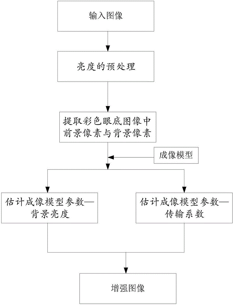
[0062] figure 1 It is a flowchart of a color fundus image image enhancement method in a specific embodiment of the present invention, specifically comprising the following steps:

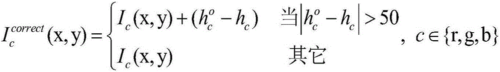
[0063] Step 1: Brightness preprocessing: record the red, green, and blue channels as R, G, and B respectively, and according to the gray value corresponding to the maximum extreme point of the gray histogram of the three channels of R, G, and B, the Color fundus image is corrected for brightness; its main purpose is to correct the overall brightness of the fundus image that is too bright and too dark.

[0064] Step 1 The specific steps include:

[0065] The brightness preprocessing of the color fundus image includes: extracting the gray histogram of the input image, setting the standard brightness, and performing brightness correction according to the standard brightness; the ...


PUM


Abstract

The invention discloses a color eye ground image enhancement method based on an imaging model. The method can improve brightness and low contrast at the same time, and realize image data fidelity, and can effectively enhance the region of interest of clinical diagnosis; and comprises the steps of preprocessing the brightness; extracting foreground pixels and background pixels in a color eye ground image: estimating imaging model parameter-background brightness and an imaging model parameter-transmission coefficient; and finally performing image enhancement according to the imaging model: substituting the background brightness and the transmission coefficient into the imaging model for calculation to obtain an enhanced image.

Description

technical field [0001] The invention belongs to the technical field of image enhancement, and in particular relates to an image enhancement method of a color fundus map based on an imaging model. Background technique [0002] The screening of fundus images is an effective means to collect retinal information of patients. Whether it is manual diagnosis or computer-aided automatic diagnosis, high-quality fundus images are very important for lesion detection and reliable diagnosis results. Due to eye diseases, imaging equipment, and patient cooperation, the obtained fundus images often have problems such as uneven brightness and low contrast. The low contrast in the fundus image will cause the characteristics of the fundus structure and lesion structure to be blurred, which will seriously affect the correct diagnosis of fundus diseases. Since the low contrast of fundus images is sometimes caused by ocular lesions, such as cataracts, image quality cannot be improved by reacquir...

Claims


Application Information

Patent Timeline
IPC IPC(8): G06T5/00G06T7/00
CPCG06T5/005G06T7/0012G06T2207/30041G06T2207/30101
Inventor 李慧琦熊荔
Owner BEIJING INSTITUTE OF TECHNOLOGYGY
Why Eureka
Try Eureka