An automatic extraction method of vertebrae in MRI lumbar spine images

A technology of automatic extraction and image processing, which is applied in the field of medical image processing to achieve rapid quantitative diagnosis, improve efficiency, and improve the efficiency of diagnosis work.

Active Publication Date: 2021-04-13
周兴祥 +2
View PDF3 Cites 0 Cited by
  • Summary
  • Abstract
  • Description
  • Claims
  • Application Information

AI Technical Summary

Problems solved by technology

This method solves the problem of automatic identification and extraction of vertebrae in MRI lumbar images, and improves the efficiency of doctors' diagnosis

Method used


Image

  • An automatic extraction method of vertebrae in MRI lumbar spine images

Examples


Embodiment Construction

[0017] The present invention will be described in further detail below according to the drawings and specific embodiments.

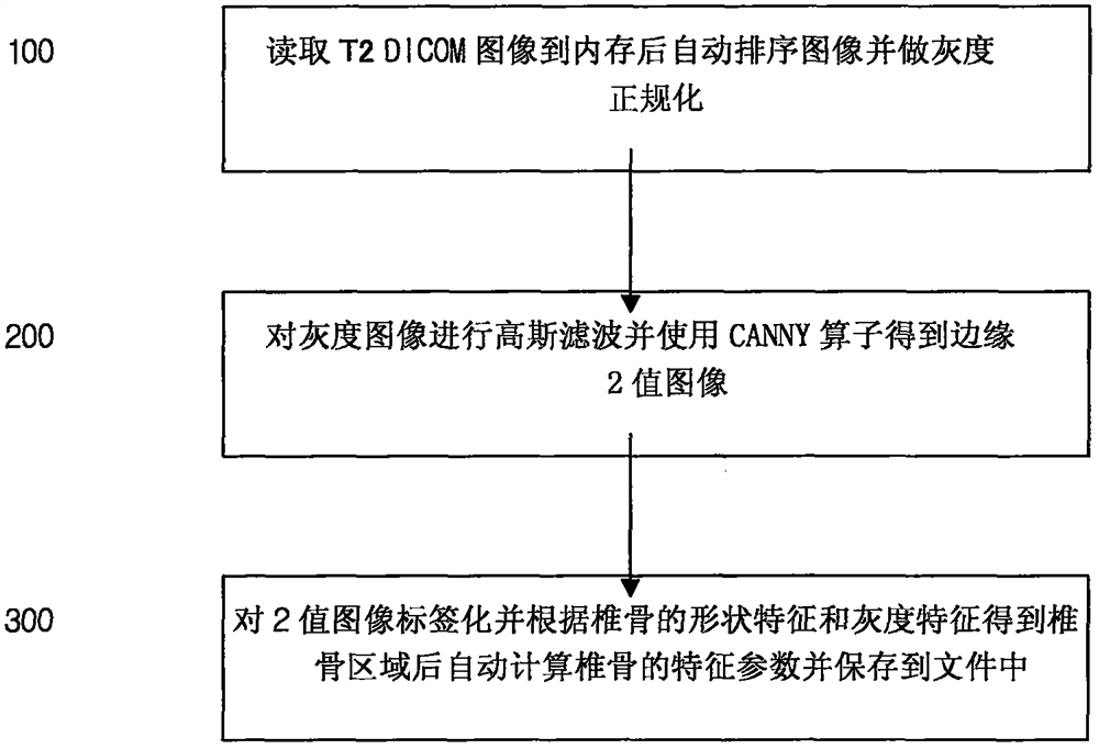
[0018] like figure 1 Shown, the automatic extraction method of vertebra in a kind of MRI lumbar vertebra image of the present invention comprises the steps:

[0019] 100: Read the T2DICOM image file of the patient's lumbar spine MRI examination in the specified folder to the memory and normalize the gray value of the image to the range of 0-255;

[0020] 200: Perform Gaussian filtering on the grayscale image and use the CANNY operator to obtain an edge binary image;

[0021] 300: Labeling the binary image and obtaining the position and number of vertebrae according to the shape and grayscale features of the vertebrae and calculating the vertebral feature quantity;

[0022] The DICOM image of the patient's lumbar spine examination obtained from the MRI diagnostic device is saved in a designated folder.

[0023] In 100, after selecting all the T2 image ...


PUM


Abstract

Invented an automatic extraction method of vertebrae in MRI lumbar images. The invention relates to the technical field of medical image processing, in particular to an automatic extraction method of vertebrae in MRI (magnetic resonance imaging) lumbar images. With the advancement of medical imaging technology, MRI has become an important means of lumbar disease diagnosis. Traditionally, it takes more than 10 minutes to locate a lumbar vertebra of a patient with a mouse, which directly affects the efficiency of doctors' diagnosis. The invention solves the problem of automatic recognition and extraction of vertebrae in MRI lumbar images. Using the T2 image of the MRI lumbar spine, the vertebrae are automatically extracted and the area, width, height, and average gray level of the vertebrae are automatically calculated, which realizes the rapid quantitative diagnosis of lumbar spine diseases and improves the efficiency of doctors' diagnosis of lumbar spine diseases.

Description

technical field [0001] The invention relates to the technical field of medical image processing, in particular to an automatic extraction method for vertebrae in MRI (magnetic resonance imaging) lumbar images. Background technique [0002] With the advancement of medical imaging technology, MRI has become an important means of lumbar disease diagnosis. In order to realize the quantitative diagnosis of diseases such as lumbar vertebral slippage and vertebral fracture, it is necessary to accurately segment and identify each vertebral body from the diagnostic image. However, it is far from enough to rely solely on manual methods to complete this work. The traditional assessment of vertebral fractures is to manually mark 6 points (the four corners and the midpoint of the upper and lower edges) for each vertebra, and then measure the heights of the front, middle, and back vertebrae. But this process is quite time-consuming. It takes more than 10 minutes to locate one vertebra ...

Claims


Application Information

Patent Timeline
Patent Type & Authority Patents(China)
IPC IPC(8): G06T7/12G06T7/62
CPCG06T2207/20182G06T2207/30012
Inventor 周兴祥于全文汤光宇
Owner 周兴祥
Why Eureka
Try Eureka