Application of anti-human CD4 and anti-human CD184 monoclonal antibodies serving as markers

A technology of monoclonal antibody and application, which is applied in the medical field and can solve problems such as high positive rate, no specific diagnostic value of CTD-ILD, unstable ELISA detection results, etc.

Active Publication Date: 2017-06-13
SHANGHAI CITY JIADING DISTRICT CENT HOSPITAL
View PDF3 Cites 2 Cited by
  • Summary
  • Abstract
  • Description
  • Claims
  • Application Information

AI Technical Summary

Problems solved by technology

[0005] At present, there is still a lack of clinical biomarkers for the diagnosis of ILD. The more specific serological marker is the detection of KL-6 by enzyme-linked immunoassay. Stable, with a high positive rate, and existing reports say that KL-6 has no significant correlation with disease activity, and has no specific diagnostic value for CTD-ILD

Method used


Image

  • Application of anti-human CD4 and anti-human CD184 monoclonal antibodies serving as markers
  • Application of anti-human CD4 and anti-human CD184 monoclonal antibodies serving as markers
  • Application of anti-human CD4 and anti-human CD184 monoclonal antibodies serving as markers

Examples


Embodiment 1

[0072] Isolation of PBMC from peripheral blood of patients

[0073] Take 5ml of heparin anticoagulant blood from the patient, mix it upside down repeatedly; dilute the blood sample with an equal amount of PBS.

[0074] Inject the diluted blood sample obliquely and slowly along the wall above 4ml of lymphatic separation solution; centrifuge at 1200g for 10min in a slow-rising and slow-falling manner.

[0075] Absorb part of the cells in the turbid zone in the centrifuge tube, transfer to another centrifuge tube added with PBS, 600g, and centrifuge for 7 minutes quickly;

[0076] Discard the supernatant, resuspend in 200 μl, count the cells, and dilute to a lymphocyte count of 0.5×10 9 / g, standby.

Embodiment 2

[0078] Preparation of mixed fluorescent antibodies

[0079] Adjust the concentration of anti-human CD4 monoclonal antibody (Abcam Company) to 15 mg / ml and anti-human CD184 monoclonal antibody (Abcam Company) to 25 mg / ml respectively, and place them in an ice bath for magnetic stirring for 10 min, without foaming during the stirring process.

[0080] FITC (Sigma Company) was weighed according to 0.01 mg / mg protein for labeling CD184.

[0081] APC (Sigma Company) was weighed according to 0.05 mg / mg protein for labeling CD4.

[0082] Slowly add fluorescein to the antibody protein solution under stirring, this step is completed within 5 minutes, place the mixed solution at 4 degrees, and continue magnetic stirring for 18 hours.

[0083]The protein mixture was taken out, centrifuged at 2500 rpm / min for 25 min, and dialyzed overnight at 4°C using buffered saline with a pH value of 8.0.

[0084] The protein mixture dialyzed overnight was passed through a glucose gel G50 column to r...

Embodiment 3

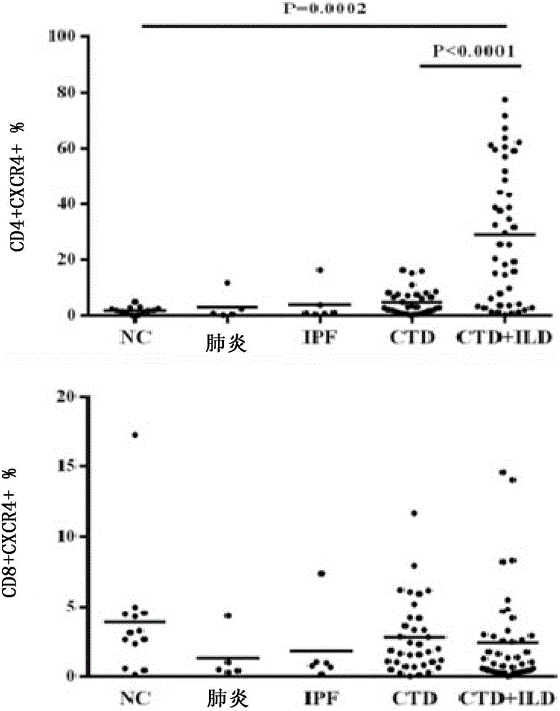
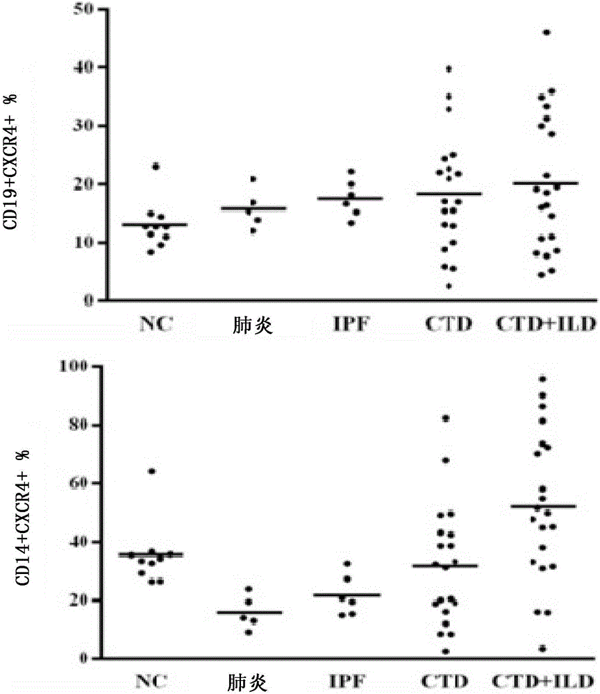
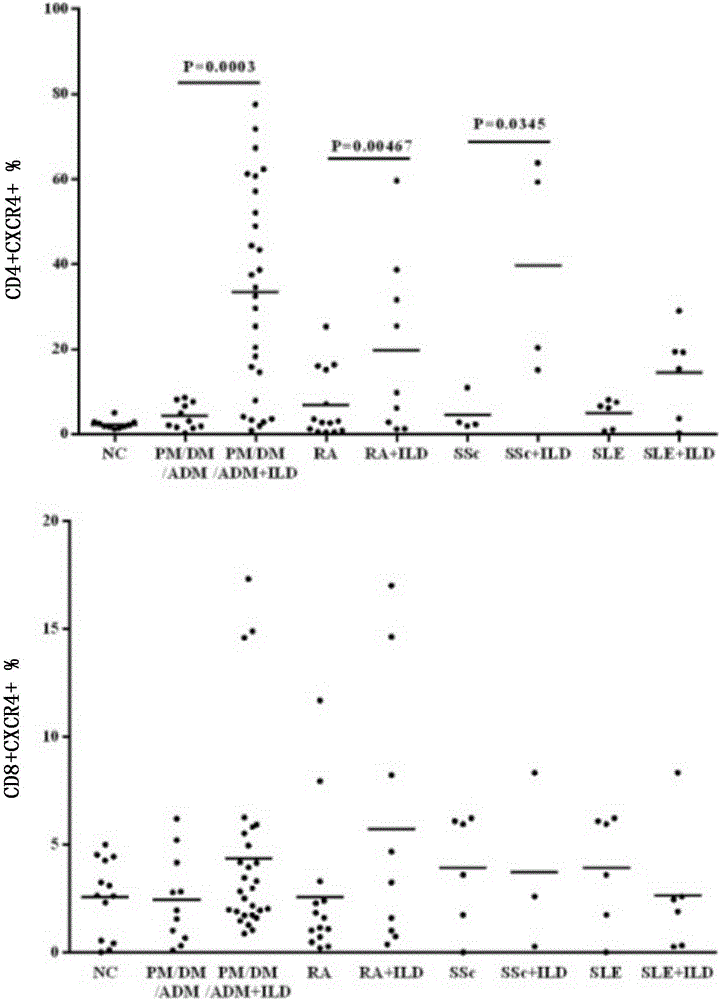
[0088] Double-antibody flow cytometry combined detection method based on the proportion of CD4+CD184+T cells in peripheral blood PBMC

[0089] Take 5ml of fresh heparin anticoagulated blood from the patient, and mix it upside down repeatedly; extract PBMC from the patient's peripheral blood.

[0090] Count the extracted PBMC with a hematology analyzer, and dilute the PBMC to 0.5×10 based on the number of lymphocytes. 9 / g.

[0091] Aspirate 100 μl of diluted PBMC, add 10 μl of mixed antibody prepared in Example 2, and incubate in the dark for 15 minutes.

[0092] Add 200μl PBS, mix well, and perform flow cytometry detection on the machine.


PUM


Abstract

The invention discloses application of an compound of an anti-human CD4 monoclonal antibody and an anti-human CD184 monoclonal antibody serving as markers, in particular relates to application of the anti-human CD4 monoclonal antibody and the anti-human CD184 monoclonal antibody serving as markers for diagnosing connective tissue disease correlated interstitial pneumonia, application of the anti-human CD4 monoclonal antibody and the anti-human CD184 monoclonal antibody to preparation of a reagent or kit for diagnosing the connective tissue disease correlated interstitial pneumonia, or application of the anti-human CD4 monoclonal antibody and the anti-human CD184 monoclonal antibody to preparation of a reagent or kit for judging the severity degree, regression or prognosis of the connective tissue disease correlated interstitial pneumonia. The invention further provides a corresponding kit.

Description

technical field [0001] The invention relates to the medical field, in particular to a marker that can be used to detect interstitial pneumonia associated with connective tissue diseases and its application. Background technique [0002] Connective tissue disease is a group of autoimmune diseases characterized by damage to its own connective tissue. Connective tissue disease (CTD) has many specific types, such as systemic lupus erythematosus (SLE), rheumatoid arthritis (RA), systemic sclerosis (SSc), Sjogren's syndrome (pSS), polymyositis And dermatomyositis (PM / DM, such as amyopathic dermatomyositis ADM), mixed connective tissue disease (MCTD), spondyloarthropathy (SpA), etc. [0003] The clinical manifestations of Connective Tissue Disease (CTD) are complex and changeable, involving multiple organs and tissues, among which pulmonary involvement is the most common, and it is also a disease with high fatality that seriously threatens the lives of patients and is one of the i...

Claims


Application Information

Patent Timeline
Patent Type & Authority Applications(China)
IPC IPC(8): G01N33/577
CPCG01N33/577
Inventor 庄磊靓
Owner SHANGHAI CITY JIADING DISTRICT CENT HOSPITAL
Why Eureka
Try Eureka