Analysis method and device for multi-mode radiomics, and terminal

A technology of radiomics and analysis methods, applied in the field of biomedical engineering, can solve the problem of inability to extract image features from multiple aspects, and achieve the effect of enriching the number of source features

Active Publication Date: 2017-06-20
SHENZHEN INST OF ADVANCED TECH CHINESE ACAD OF SCI
View PDF3 Cites 10 Cited by
  • Summary
  • Abstract
  • Description
  • Claims
  • Application Information

AI Technical Summary

Problems solved by technology

[0003] In view of this, the embodiment of the present invention provides a multimodal radiomics analysis method, device and terminal to solve the problem that the prior art cannot extract image features in multiple aspects in radiomics research, so as to maximize Mining medical image information

Method used


Image

  • Analysis method and device for multi-mode radiomics, and terminal
  • Analysis method and device for multi-mode radiomics, and terminal
  • Analysis method and device for multi-mode radiomics, and terminal

Examples


Embodiment Construction

[0048] In order to make the object, technical solution and advantages of the present invention clearer, the present invention will be further described in detail below in conjunction with the accompanying drawings and embodiments. It should be understood that the specific embodiments described here are only used to explain the present invention, not to limit the present invention.

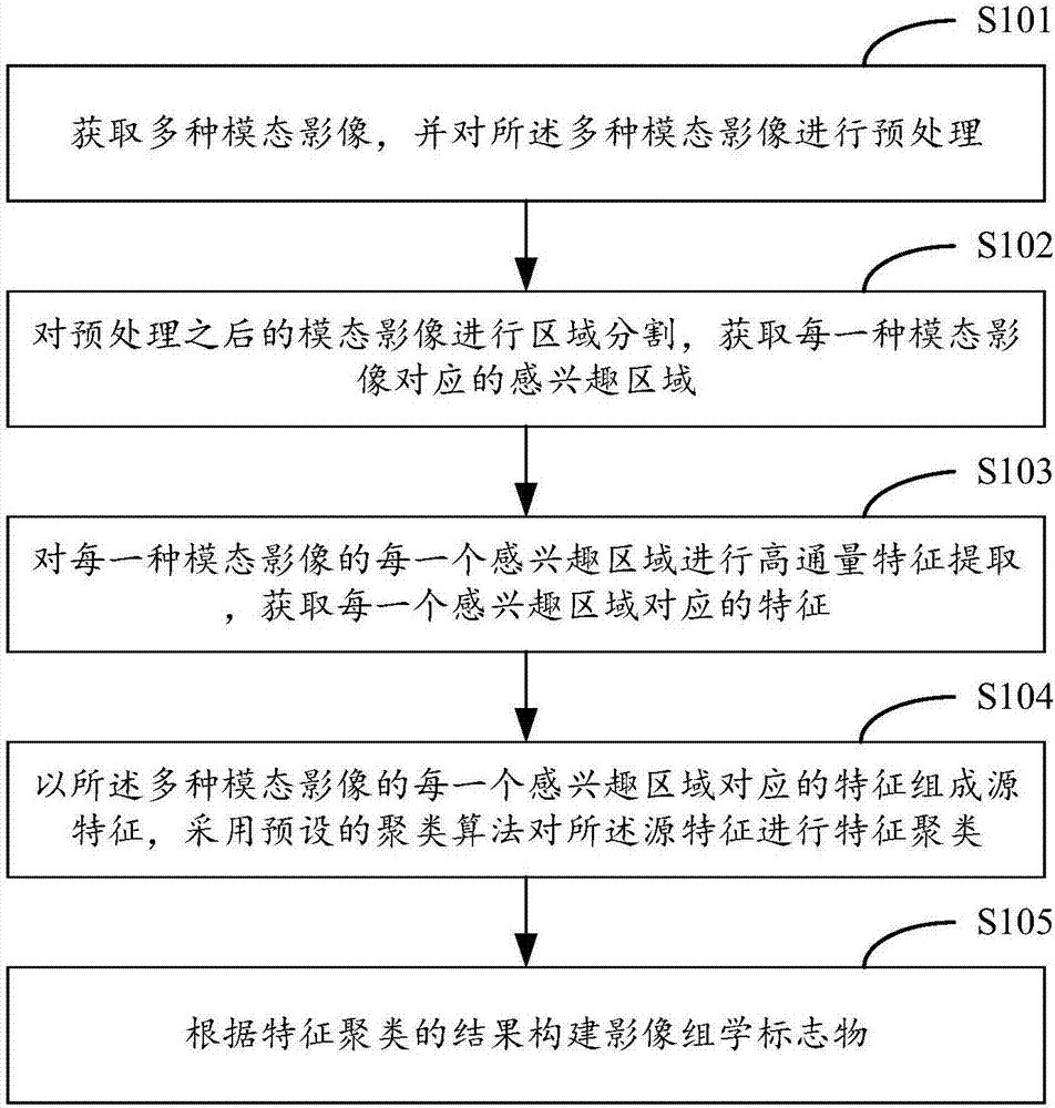
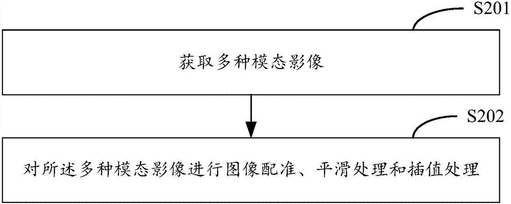
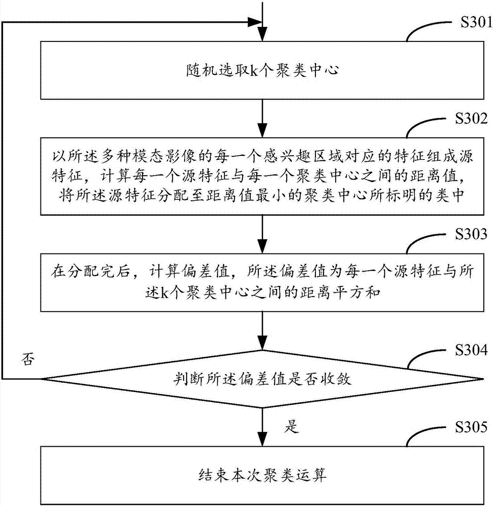
[0049] In the embodiment of the present invention, multiple modality images are obtained, and the multiple modality images are preprocessed; and then the preprocessed modality images are segmented to obtain the region of interest corresponding to each modality image ; Carry out high-throughput feature extraction for each region of interest of each modality image, and obtain the corresponding features of each region of interest; finally use the feature composition corresponding to each region of interest of the multiple modality images Source features, using a preset clustering algorithm to perform ...


PUM


Abstract

The invention provides an analysis method and device for multi-mode radiomics, and a terminal. The method comprises steps of obtaining multiple mode images, and preprocessing the multiple mode images; performing regional segmentation to the preprocessed mode images to obtain an area of interest corresponding to each mode image; for each area of interest of each mode image, performing high-pass feature extraction, and obtaining characteristics corresponding to each area of interest; grouping the characteristics corresponding to each area of interest of the multiple mode images into source characteristics, and using a preset clustering algorithm to perform characteristic clustering of the source characteristics; and according to the result of the characteristic clustering, constructing radiomics markers. Therefore, the problem that image characteristics cannot be extracted from multiple aspects in the radiomics research in the prior art is solved, the number of the source characteristics for the characteristic clustering and the representative characteristic kind after clustering are enriched, and the medical image information can be excavated to the greatest extent.

Description

technical field [0001] The invention belongs to the technical field of biomedical engineering, and in particular relates to an analysis method, device and terminal of multimodal imaging omics. Background technique [0002] Traditional medical image analysis generally only focuses on the manifestation of tumors in a single modality. Radiomics can transform traditional medical images into mineable data information. In the research of radiomics, the existing technology mainly obtains the static information of four anatomical images, without considering the dynamic information contained in the images, and cannot extract image features in many ways, resulting in the number of extracted features and sample cases Limited, unable to maximize the mining of medical image information. Contents of the invention [0003] In view of this, the embodiment of the present invention provides a multimodal radiomics analysis method, device and terminal to solve the problem that the prior art...

Claims


Application Information

Patent Timeline
Patent Type & Authority Applications(China)
IPC IPC(8): G06T7/11G06T7/32
Inventor 宋柏霖李志成李其花孙秋畅王梦巧
Owner SHENZHEN INST OF ADVANCED TECH CHINESE ACAD OF SCI
Why Eureka
Try Eureka