
Automatic region-of-interest segmentation and registration of dynamic contrast-enhanced images of colorectal tumors

A dynamic contrast enhancement, image technology

Inactive Publication Date: 2017-08-01
AGENCY FOR SCI TECH & RES
View PDF4 Cites 3 Cited by
  • Summary
  • Abstract
  • Description
  • Claims
  • Application Information

AI Technical Summary

Problems solved by technology

This usually requires the user to manually outline the region of interest on multiple (usually around 50 or more) DCE images, which is time-consuming and tedious

Method used


Image

  • Automatic region-of-interest segmentation and registration of dynamic contrast-enhanced images of colorectal tumors
  • Automatic region-of-interest segmentation and registration of dynamic contrast-enhanced images of colorectal tumors
  • Automatic region-of-interest segmentation and registration of dynamic contrast-enhanced images of colorectal tumors

Examples


Embodiment Construction

[0029] It should be further appreciated that the exemplary embodiments are exemplary only, and are not intended to limit the scope, applicability, operation or configuration of the invention in any way. Rather, the foregoing detailed description will provide those skilled in the art with a convenient road map for implementing the exemplary embodiments of the invention, and it should be understood that other modifications may be made in the exemplary embodiments without departing from the scope of the invention as defined in the appended claims. Various changes have been made in terms of the functions and arrangement of elements and methods of operation described in the exemplary embodiments.

[0030] Some portions of the description below are presented explicitly or implicitly in terms of algorithms and functional or symbolic representations of operations on data in computer memory. These algorithmic descriptions and functional or symbolic representations are the means used by...


PUM


Abstract

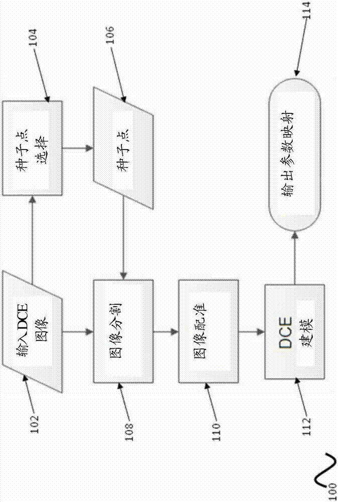
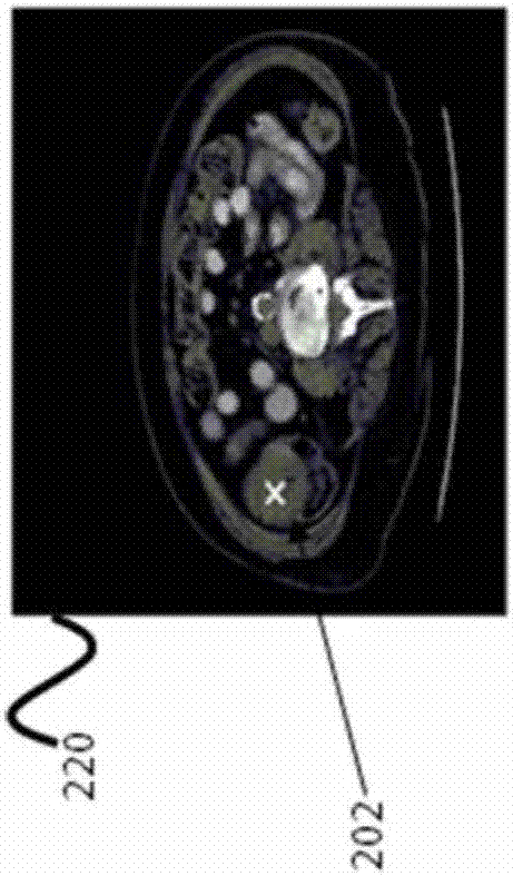
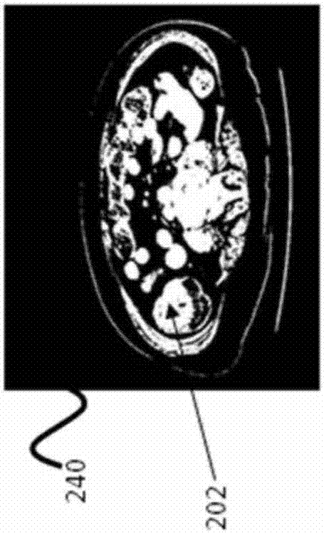
A method for dynamic contrast enhanced (DCE) image processing and kinetic modeling of an organ's region-of-interest is provided. The method includes deriving at least a contour of an exterior of the organ's region-of-interest from one or more of a plurality of images; generating a spline function in response to the derived contour of the exterior of the organ's region-of-interest from the one or more of the plurality of images; registering the plurality of images wherein the organ's region-of-interest has been segmented; deriving a tracer curve for the organ's region-of-interest in the registered images, the tracer curve indicating a change in concentration of a contrast agent flowing through the organ's region-of-interest over a time period; and kinetic modeling by fitting a kinetic model to the tracer curve to generate one or more maps of tissue physiological parameters associated with the kinetic model.

Description

technical field [0001] The present invention relates to the processing of image sequences produced by dynamic contrast enhanced (DCE) imaging for the diagnosis of neoplastic diseases. In particular, the present invention relates to pre-DCE modeling for segmentation and registration of organ tissues of interest. Background technique [0002] An important aspect of biomedical-related research is the detection and analysis of tumors in organs. With current technology, images of organs are often manually analyzed to detect the presence of tumors. However, manually analyzing images is time-consuming and tedious. [0003] One commonly used conventional technique is Magnetic Resonance Imaging (MRI). MRI technology is widely used for imaging soft tissues within the human (or animal) body, and there is much work in developing techniques to perform analysis in such a way that the imaged tissue is characterized, for example, as normal or diseased. However, conventional MRI has so f...

Claims


Application Information

Patent Timeline
Patent Type & Authority Applications(China)
IPC IPC(8): G06T7/12G06T7/73G01R33/56G06V10/40
CPCG01R33/5608G06T2207/10096G06T2207/20101G06T2207/30028G06T2207/30096G06T7/73G06T7/12G06T7/33G06T7/246G06V10/40G06T7/0012
Inventor 侯祖军汪悦
Owner AGENCY FOR SCI TECH & RES
Learn More