
Medical image file multi-plane processing method and device

A medical image and processing method technology, which is applied in the field of medical image file multi-plane processing method and device thereof, can solve the problems of new medical cost, does not include MIP and the like, and achieves the effect of convenient medical diagnosis

Active Publication Date: 2017-08-11
BEIJING ALLCURE MEDICAL TECH CO LTD
View PDF4 Cites 0 Cited by
  • Summary
  • Abstract
  • Description
  • Claims
  • Application Information

AI Technical Summary

Problems solved by technology

The existing multi-plane reconstruction process in the medical field does not include the MIP function. Many hospitals still cannot meet the needs after using MPR, and need to purchase a separate MIP module, which increases medical costs.

Method used


Image

  • Medical image file multi-plane processing method and device
  • Medical image file multi-plane processing method and device
  • Medical image file multi-plane processing method and device

Examples


Embodiment Construction

[0042] The specific implementation of the method for multi-plane processing of medical image files provided by the embodiments of the present invention will be described below with reference to the accompanying drawings.

[0043] 1. Method embodiment

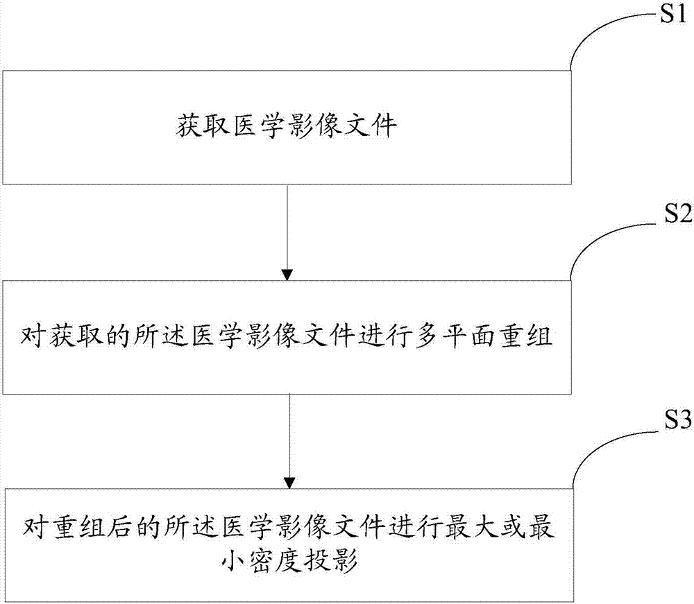
[0044] The medical image file multi-plane processing method described in this embodiment, such as figure 1 shown, including the following steps:

[0045] S1. Obtain a medical image file; the medical image file is an image format file conforming to the international "Medical Digital Imaging and Communication" standard, preferably, the medical image file is a dicom image format file;

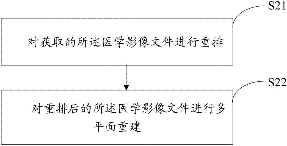
[0046] S2. Perform multi-plane reorganization on the acquired medical image file; superimpose all the axial images within the scanning range, and carry out coronal, sagittal, and oblique at any angle on the tissue designated by the reorganization line marked by certain marking lines. Bit image reorganization.

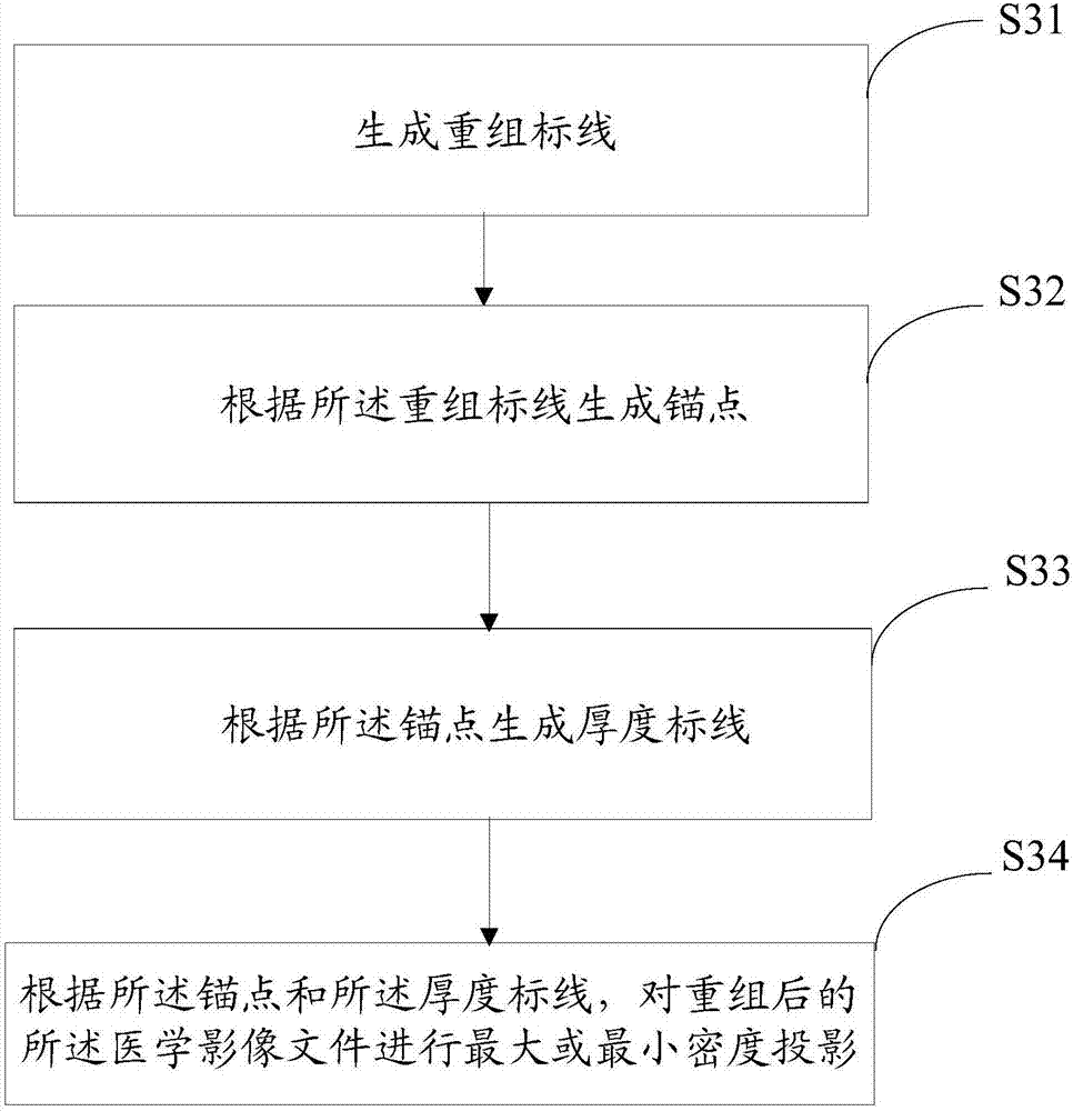
[0047] S3. Perform maximum or minim...


PUM


Abstract

The method provides a medical image file multi-plane processing method. The method comprises steps of accessing to a medical image file; subjecting the obtained medical image file to multi-plane reconstruction; and performing the maximum intensity projection or minimum intensity projection of the medical image file after reconstruction. The invention also discloses a medical image file multi-plane processing device. The processing method and the processing device can not only reconstruct the coronal, sagittal and arbitrary angle oblique images of the specified tissue, but also can be used for maximum or minimum intensity projection of relatively high density tissues and structures such as blood vessels, bones, lung masses and significantly enhanced soft tissue focuses, which greatly facilitates medical diagnosis.

Description

technical field [0001] The invention relates to medical image communication and transmission technology, in particular to a multi-plane processing method and device for medical image files. Background technique [0002] Multi-planar reconstruction (MPR, multi-planner reformation) is to superimpose all the axial images within the scanning range and then perform coronal, sagittal, and oblique image reconstruction at any angle on the tissue designated by the reorganization line marked by certain reticles . A new tomographic image can be generated arbitrarily without repeated scanning. [0003] The maximum and minimum intensity projection (MIP, maximum / minimum intensity projection) is to project the maximum or minimum density in the selected tissue layer in a certain plane direction. MIP is widely used in the diagnosis of relatively high-density tissues and structures, such as blood vessels, bones, lung masses, and obviously enhancing soft tissue lesions. The existing multi-p...

Claims


Application Information

Patent Timeline
Patent Type & Authority Applications(China)
IPC IPC(8): G06T15/08
CPCG06T15/08G06T2210/41
Inventor 李益杰康世功阎俊
Owner BEIJING ALLCURE MEDICAL TECH CO LTD
Learn More