Defining scanning parameters of a ct scan using external image capture

A technology of CT scanning and scanning parameters, applied in medical images, computed tomography scanners, instruments used for radiological diagnosis, etc., can solve problems such as longer waiting time, and achieve the effect of reducing time length, radiation load, and widespread use

Active Publication Date: 2017-11-10
SIEMENS HEALTHCARE GMBH
View PDF9 Cites 9 Cited by
  • Summary
  • Abstract
  • Description
  • Claims
  • Application Information

AI Technical Summary

Problems solved by technology

[0010] However, the planning of CT imaging sequences based on internally stored information positional representations implies an additional workload
In addition, the patient must wait longer in the CT system to be able to obtain the location map of the stored information

Method used


Image

  • Defining scanning parameters of a ct scan using external image capture
  • Defining scanning parameters of a ct scan using external image capture
  • Defining scanning parameters of a ct scan using external image capture

Examples


Embodiment Construction

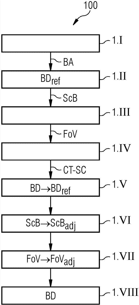
[0055] figure 1 A flowchart 100 is shown illustrating an exemplary embodiment of a method of using a CT system to define scan parameters for a CT scan of a region of interest of an examination subject. In step 1.I, image capture BA of the patient is initially performed using a camera. The camera is arranged relative to the examination object (in this case the patient) such that at least a segment of the patient covering a desired field of view FoV can be recorded. exist figure 1 In the exemplary embodiment shown, the camera used is a 3D camera that acquires a 3D profile of the patient. In step 1.II, based on the captured image BA, CT reference image data BD that matches the captured image BA as closely as possible is obtained from the database ref. For example, the size of the body region recorded on the captured image BA must be the same as the reference image data BD ref The corresponding size matches. Based on the selected CT reference image data BD ref , to determin...


PUM


Abstract

A method for defining scanning parameters of a CT scan of a region of interest of an examination object (O) using a CT system is described. In an embodiment of the method, external image capture of external features of the examination object is carried out using an additional image capture unit. In addition, at least one scanning parameter in the axial direction of the CT system is determined on the basis of the external image capture. Finally, a CT scan is performed using the at least one scanning parameter determined. A CT scan range defining device is additionally described. A computed tomography system is also described.

Description

technical field [0001] The present invention relates to a method for defining scanning parameters of a CT scan of a region of interest of an examination object using a CT system. The invention also relates to a CT scanning parameter defining device. The invention finally relates to a computer tomography system. Background technique [0002] With modern imaging methods, two-dimensional or three-dimensional image data are often generated, which can be used to visualize the examination object, but also for other applications. [0003] Imaging methods are generally based on the detection of X-radiation, wherein so-called projection measurement data are generated. The projection measurement data may be acquired, for example, using a computed tomography system (CT system). In a CT system, the gantry-mounted combination of an X-ray source and an oppositely disposed X-ray detector will generally rotate around a scanning room in which an examination object (which, without limitati...

Claims


Application Information

Patent Timeline
Patent Type & Authority Applications(China)
IPC IPC(8): A61B6/03
CPCA61B6/032A61B6/481A61B6/54A61B6/545A61B6/488A61B6/5205A61B6/542G16H30/20G16H40/63
Inventor U·福伊尔莱因S·格罗斯科普夫G·索泽M·聚林
Owner SIEMENS HEALTHCARE GMBH
Why Eureka
Try Eureka