
Method for estimating time of occurrence of infarct region on basis of brain image

A technology of brain image and region, applied in the field of estimating the occurrence time of infarction region based on brain image, which can solve the problems of low objectivity and reliability

Active Publication Date: 2017-12-01
THE ASAN FOUND
View PDF6 Cites 4 Cited by
  • Summary
  • Abstract
  • Description
  • Claims
  • Application Information

AI Technical Summary

Problems solved by technology

In addition, although the luminance in the FLAIR image that changes over time is displayed as a spectral distribution, when the timing of occurrence is determined by a specific SIR value limit, the objectivity and reliability are low

Method used


Image

  • Method for estimating time of occurrence of infarct region on basis of brain image
  • Method for estimating time of occurrence of infarct region on basis of brain image
  • Method for estimating time of occurrence of infarct region on basis of brain image

Examples


Embodiment Construction

[0029] Hereinafter, the present disclosure will now be described in detail with reference to the accompanying drawing(s) with reference to the accompanying drawings.

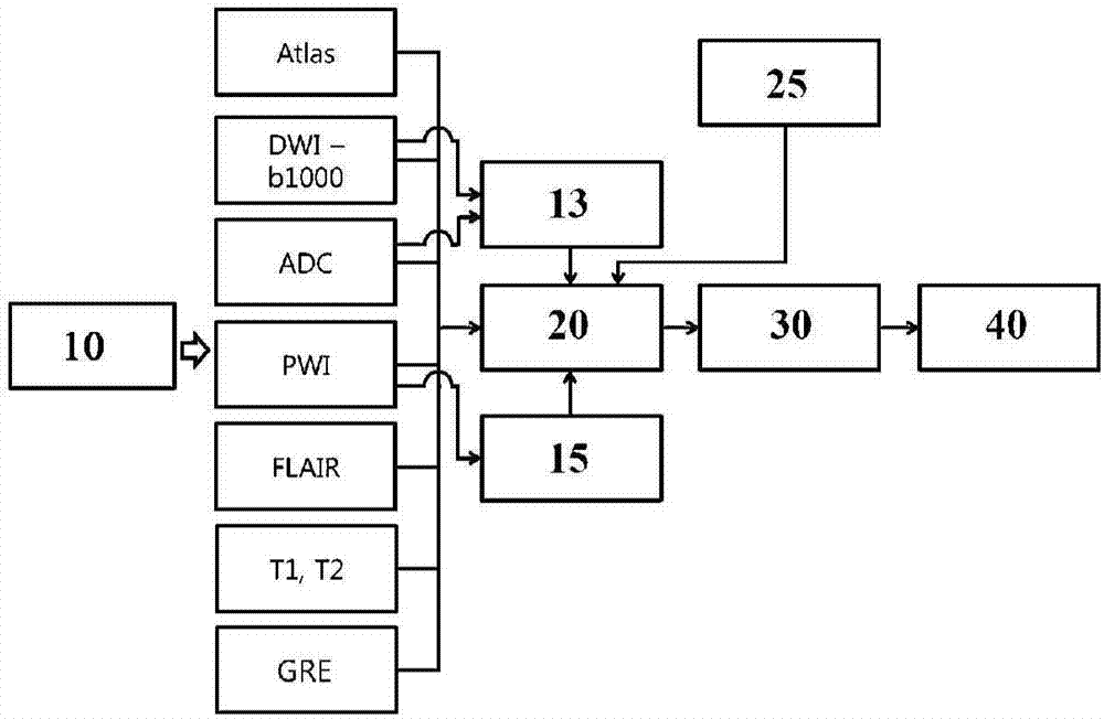
[0030] figure 1 It is a diagram explaining an example of the method of estimating the occurrence time of the infarct area based on the brain image of the present disclosure.

[0031] In the method of estimating the occurrence time of the infarct area based on the brain image, the brain image (10) is obtained. From the infarct region (infarct region) included in the obtained brain image (for example: DWI, ADC, PWI, FLAIR, T1, T2, etc.), a quantitative value that changes according to the onset time of the infarct region is extracted ( quantitative value) set (30). After that, a prepared correspondence relation (prepared) is used so that the quantitative value set corresponds to the occurrence time point of the infarct area, so that the quantitative value set corresponds to the occurrence time point of the infarct area...


PUM


Abstract

The disclosure relates to a method for estimating the time of occurrence of an infarct region based on basis of a brain image, including: a step of obtaining a brain image; a step of extracting a fixed value set that changes according to an onset time of the infarcted region in the brain image; and a step of using corresponding relation prepared such that the fixed value set corresponds to the onset time of the infarct region, so that the fixed value set corresponds to the onset time of the infarction region.

Description

Technical field [0001] The present disclosure (Disclosure) as a whole relates to a method of estimating the time point of occurrence of an infarct region based on brain images (METHOD FOR ESTIMATING ONSET TIME OF INFARCT REGION BASED ON BRAIN IMAGE), and in particular, relates to the use of unknown information A method of estimating the occurrence time of the infarct region based on the brain image by integrating quantitative values ​​extracted from the brain image. Background technique [0002] Here, the provision of background technology of the present invention does not mean that these are all known technologies (This section provides background information related to the present disclosure which is notnecessaril y prior art). [0003] Recently, a method of evaluating the state of a lesion using medical images has been used. For example, in the case of acute stroke patients such as cerebral infarct (cerebralinfarct) or cerebral hemorrhage (cerebral hemorrhage), brain images are...

Claims


Application Information

Patent Timeline
Patent Type & Authority Applications(China)
IPC IPC(8): A61B5/055A61B5/00
CPCA61B5/00A61B5/055G06T7/0012G06T7/30A61B5/0042G01R33/5608G01R33/56341G01R33/56366G06T7/0014G06T2207/10088G06T2207/30016
Inventor 金南局姜东和
Owner THE ASAN FOUND
Learn More