Cone-beam computed tomography image and X-ray image registration method

A tomography, optical image technology, applied in the field of oral clinical medicine and computer vision, can solve problems such as non-rigid and complex structural changes in three-dimensional craniofacial images that are difficult to process

Active Publication Date: 2017-12-22
PEKING UNIV
View PDF5 Cites 31 Cited by
  • Summary
  • Abstract
  • Description
  • Claims
  • Application Information

AI Technical Summary

Problems solved by technology

The method based on convolutional neural network is used for 2D and 3D image registration, but the existing registration work based on convolutional neural network regression model only deals with

Method used


Image

  • Cone-beam computed tomography image and X-ray image registration method
  • Cone-beam computed tomography image and X-ray image registration method
  • Cone-beam computed tomography image and X-ray image registration method

Examples


Embodiment Construction

[0052] Below in conjunction with accompanying drawing, further describe the present invention through embodiment, but do not limit the scope of the present invention in any way.

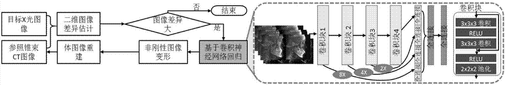
[0053] The convolutional neural network regression-based method provided by the present invention performs non-rigid registration between two-dimensional X-ray images and three-dimensional cone-beam CT images. A convolutional neural network-based regression model was used to establish the correlation between non-rigid deformation parameters of 2D X-ray images and 3D cone-beam CT images. Combining a regression model based on a hybrid residual convolutional neural network and an iterative optimization mechanism for deformation parameters, reliable online 2D and 3D image registration is achieved.

[0054] figure 1 It is a block flow diagram of the method of the present invention. The present invention will be further described below with reference to the accompanying drawings.

[0055] Step 1: Extrac...


PUM


Abstract

The invention discloses a cone-beam computed tomography image and X-ray image registration method; the method uses a regression model based on a mixing residual error convolution nerve network to build associations between two-dimension X-ray images and three dimensional cone-beam CT image non-rigid deformation parameters, and uses a mixing residual error convolution nerve network and deformation parameter iteration optimization method to realize reliable online two-dimension three dimensional image registration; the method comprises the following steps: extracting to obtain an image channel; training the regression model based on the mixing residual error convolution nerve network; carrying out two-dimension three dimensional non-rigid registration based on regression; iterating and optimizing deformation parameters; obtaining a final body image determined by the deformation parameters, and thus realizing two-dimension three dimensional image registration based on iteration regression. The method can realize reliable online two-dimension three dimensional image registration, can be applied to oral cavity clinics, and can evaluate treatments and analyze craniofacial growth according to two-dimension three dimensional image registrations.

Description

technical field [0001] The invention relates to the technical fields of oral clinical medicine and computer vision, in particular to a method for registering cone-beam computed tomography images and X-ray images. Background technique [0002] Orthodontic treatment usually lasts for several years, and multiple images collected during treatment will be used to evaluate the difference between rigid and non-rigid shapes of maxillofacial structures, especially for minor patients. Nonrigid morphological change to structural growth. Before the widespread use of cone-beam computed tomography (cone-beam CT) images in clinical orthodontics, two-dimensional radiographs were the only medium to record changes in maxillofacial morphology. Nonrigid 2D-3D image registration is a key step in obtaining 3D images from in-treatment X-ray images. Traditional 2D and 3D image registration methods rely on iterative optimization to minimize the difference between digitally-reconstructed-radiograph...

Claims


Application Information

Patent Timeline
IPC IPC(8): G06T7/33
CPCG06T7/344G06T2207/10081G06T2207/10124G06T2207/20081G06T2207/20084
Inventor 裴玉茹秦海芳张云庚郭玉珂许天民查红彬
Owner PEKING UNIV
Why Eureka
Try Eureka