Multimode medical image fusion method based on translation constant shear wave transformation

A translation-invariant, medical image technology, applied in the field of medical image processing and applications, can solve the problems of high-frequency sub-band correlation loss in different modal images

Inactive Publication Date: 2013-04-17
SOUTH CHINA UNIV OF TECH
View PDF4 Cites 34 Cited by
  • Summary
  • Abstract
  • Description
  • Claims
  • Application Information

AI Technical Summary

Problems solved by technology

Therefore, traditional fusion rules do not fully consider this correlation, resulting in the loss of high-frequency sub-band correlations of images of different modalities

Method used


Image

  • Multimode medical image fusion method based on translation constant shear wave transformation
  • Multimode medical image fusion method based on translation constant shear wave transformation
  • Multimode medical image fusion method based on translation constant shear wave transformation

Examples


Embodiment 1

[0075] Example 1: MRI-SPECT Fusion

[0076] The image fusion method of this embodiment first considers how to obtain more direction information from the original image: the decomposition of high-frequency subbands and the maintenance of translation invariance; secondly, how to combine the extracted low-frequency sub-band and high-frequency sub-band information As much as possible is transferred to the fusion image: low-frequency sub-band fusion avoids reducing the contrast of the image; high-frequency sub-band fusion avoids the loss of relevant information between sub-bands and the intervention of too much artificial experience.

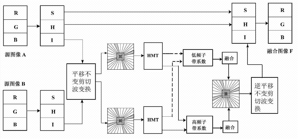
[0077] Such as figure 1 shown, including the following steps:

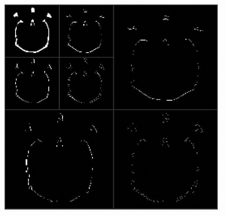
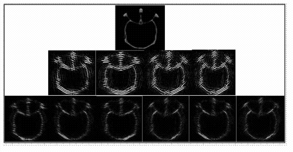
[0078] Step 1: Yes Figure 5a and Figure 5b The shown SPECT image and MRI image to be fused are subjected to IHS transformation to obtain the Intensity component of the image, and then translation-invariant shearlet decomposition is performed on it. The implementation consists of tw...

Embodiment 2

[0115] Example 2: MRI-PET Fusion

[0116] The method provided by the present invention can realize high-quality fusion of head MRI images and SPECT images under common hardware conditions, and the original resolution of MRI images and PET images is 256×256, such as Figure 7a and Figure 7b shown.

[0117] Step 1: Perform IHS transformation on the MRI image and PET image to be fused to obtain the Intensity component of the image, and then perform translation-invariant shearlet decomposition on it. The implementation consists of two steps: multi-scale segmentation and orientation localization. Whole process is with embodiment 1.

[0118] Step 2: Low-frequency approximate image fusion and high-frequency image sub-band fusion in all directions:

[0119] 1) For low-frequency approximate image fusion, a fusion rule based on the absolute value and weight of the region coefficient is adopted;

[0120] 2) For each direction subband of the high-frequency detail image, the fusion r...


PUM


Abstract

The invention discloses a multimode medical image fusion method based on translation constant shear wave transformation. The method comprises the following steps of 1) preparing two multimode medical images to be fused, utilizing translation constant shear wave transformation to respectively decompose the two images into low frequency approximation images and high frequency detail images, and decomposing the high frequency detail images into different direction sub-bands; 2) respectively fusing the sub-bands from various directions decomposed by the low frequency approximation images and the high frequency detail images of the two images; 3) carrying out inverse translation shear wave transformation on the sub-bands from various directions decomposed by the low frequency approximation images and the high frequency detail images which are fused in the step 2) to obtain a fused image. According to the method provided by the invention, as the image fusion method based on the translation constant shear wave transformation is adopted, the fusion of multimode medical image data can be realized conveniently, economically and efficiently; and the shape information and function information of detail parts implied in the internal parts of images with different modes can be fully displayed and captured, thereby meeting the accuracy requirement in medical use.

Description

technical field [0001] The invention relates to an image fusion method, in particular to a translation-invariant shearlet transform-based multi-modal medical image fusion method, which belongs to the technical field of medical image processing and application. Background technique [0002] Multimodal medical image fusion plays an important role in clinical diagnosis, and it is widely used in image-guided surgery, image-guided radiotherapy, non-invasive diagnosis and treatment planning. For example, in non-invasive diagnosis and treatment planning, doctors often need to use medical images of different modalities to see the lesion structure and details in the patient's body to accurately determine the relative position of the tumor and observe the pathology of the tumor to radioactive rays. reaction. Therefore, multimodal medical image fusion technology is a key link in modern medical visualization technology, and is widely used in modern medical clinical diagnosis. [0003]...

Claims


Application Information

Patent Timeline
Patent Type & Authority Applications(China)
IPC IPC(8): G06T5/50
Inventor 李彬王雷田联房
Owner SOUTH CHINA UNIV OF TECH
Why Eureka
Try Eureka