
Anti-radiation auxiliary device for imaging examination

An auxiliary device and radiation protection technology, which is applied in radiation safety devices, equipment for radiological diagnosis, medical science, etc., can solve the problems of insufficient radiation protection effect, insufficient effect, and poor structural flexibility, so as to promote the prevention of radiation. Radiation effect, enhanced fixing stability, and easy disassembly and assembly effects

Active Publication Date: 2018-02-09
THE AFFILIATED HOSPITAL OF SOUTHWEST MEDICAL UNIV
View PDF6 Cites 1 Cited by
  • Summary
  • Abstract
  • Description
  • Claims
  • Application Information

AI Technical Summary

Problems solved by technology

Although it plays the role of exposing the inspection area and blocking the non-inspection area, its effect is insufficient: the area of ​​the rectangular block is fixed, set S, and the rectangular block needs to be removed, then the area exposed each time is nS, where n is the number of rectangular blocks to remove, and the value of n must be at least 1
It can be seen that it has the disadvantages of poor structural flexibility and insufficient radiation protection effect.

Method used


Image

  • Anti-radiation auxiliary device for imaging examination
  • Anti-radiation auxiliary device for imaging examination
  • Anti-radiation auxiliary device for imaging examination

Examples


Embodiment

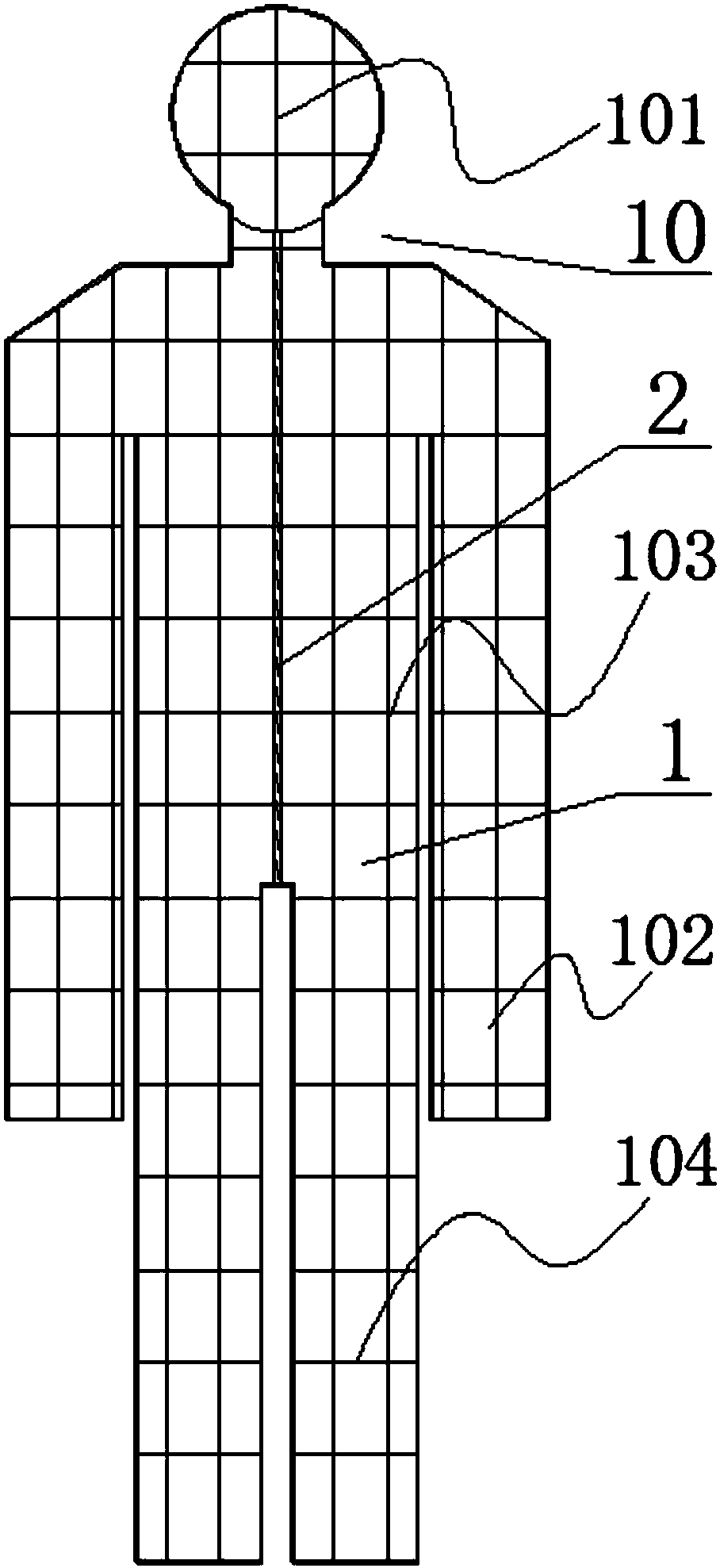
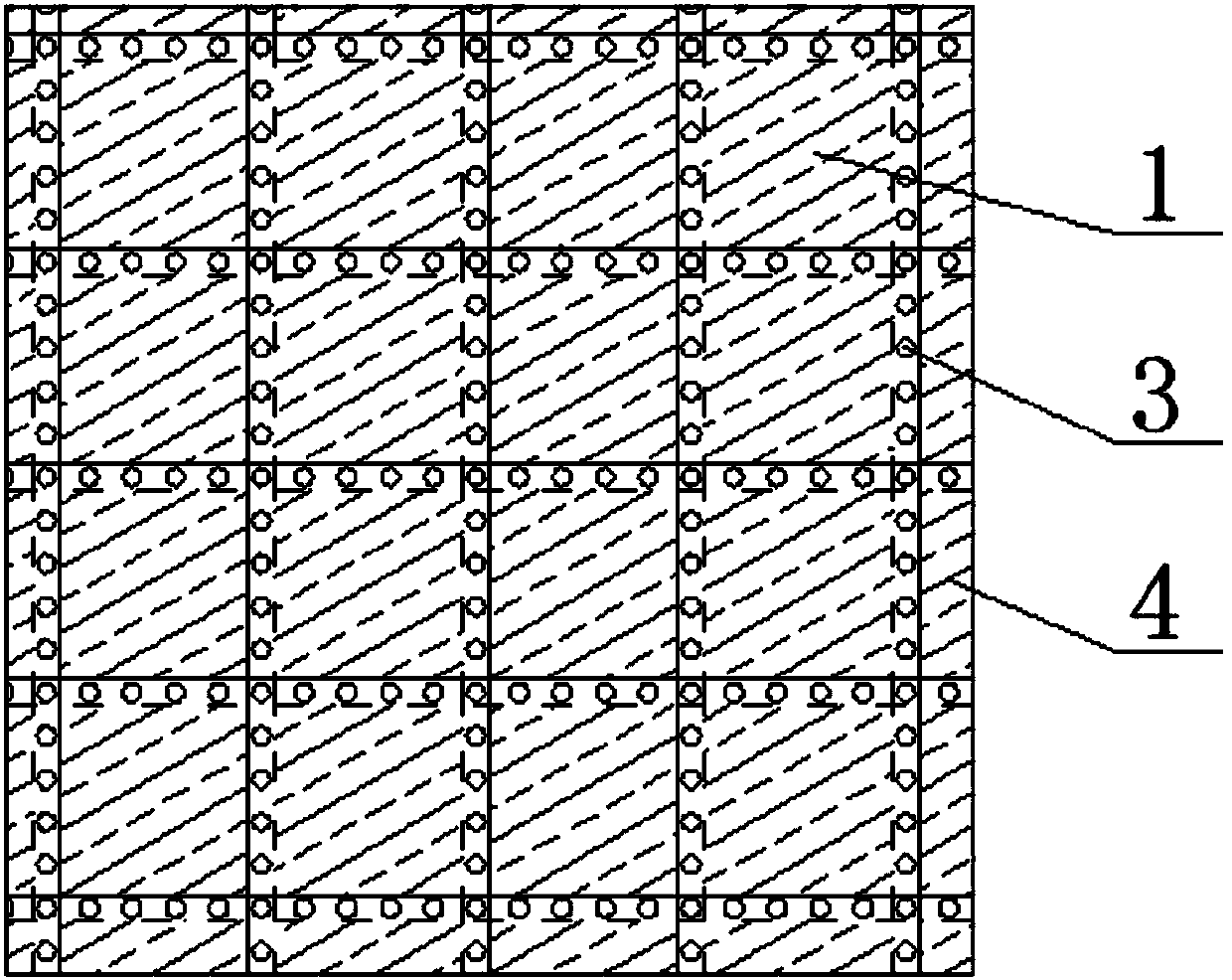
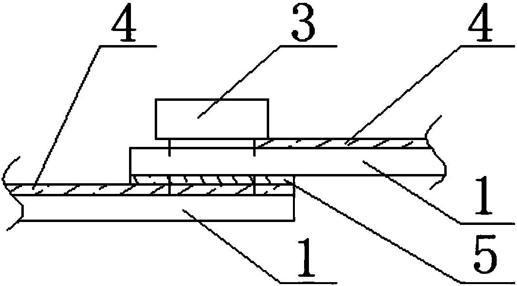
[0031] Such as figure 1 and figure 2 The radiation protection auxiliary device for image examination shown is a protective suit 10 worn by a patient when undergoing an image examination. Zippers2 that allow patients to wear radiation-resistant clothing, such as Figure 4 As shown, the material of the square cloth 1 includes a cotton layer 11 and an anti-radiation layer 12 attached to the upper surface of the cotton layer, and the anti-radiation layer 12 is equipped with a radiation-proof material inside; as image 3 As shown, the front side of the square cloth 1 is provided with Velcro-4, and the edges of each adjacent two square cloths 1 are overlapped and connected together by buttons 3, and the overlapped two square cloths 1 The overlap of the back of the square cloth 1 above is provided with Velcro 2 5; the protective clothing also includes an auxiliary attachment bag 6, such as Figure 5 and Figure 7 As shown, the auxiliary attachment bag 6 is a ring-shaped square c...


PUM


Abstract

The invention discloses an anti-radiation auxiliary device for imaging examination and belongs to the field of medical assistive devices. The device comprises anti-radiation cloth which is spliced bymultiple pieces of small square cloth and a zipper which allows a patient to wear, hook-and-loop fasteners are arranged on the front surface of the small square cloth, and every two adjacent small square cloth are connected together through multiple buttons and the hook-and-loop fasteners; the device further comprises an auxiliary attachment bag, a lead powder bag is arranged in the auxiliary attachment bag, and hook-and-loop fasteners are arranged on the back surface of the auxiliary attachment bag; a radiation opening is formed in the center of the auxiliary attachment bag, width adjustmentmechanisms are arranged on four edges of the radiation opening respectively, the width adjustment mechanisms comprise sleeves which are fixedly connected with the edges of the radiation opening, piston rods which are connected into the sleeves in a threaded rod, and shaft sleeves which sleeve the other end of each piston rod, and hook-and-loop fasteners are fixed to the edges of the radiation opening corresponding to the piston rods. The anti-radiation auxiliary device can adapt to examination parts at random positions with random sizes, can also conduct readjustment on exposed areas, can achieve relatively complete and effective anti-radiation protection, is applied conveniently, and is good in anti-radiation protection effect.

Description

technical field [0001] The invention relates to the field of medical aids, in particular to a radiation protection auxiliary device for image inspection. Background technique [0002] Imaging is a discipline in medicine. With the rapid development of computer technology, the traditional radiology department has developed into today's medical imaging department, which generally includes five inspections: X-ray, CT, magnetic resonance, ultrasound and nuclear medicine. Imaging not only expands the scope of human body examination and improves the level of diagnosis, but also can treat certain diseases. In this way, the work content of the imaging discipline has been greatly expanded, and it has become an important pillar of medical work. Not long after Wilhelm Conrad Rontgen discovered X-rays in 1895, in medicine, X-rays have been used to examine the human body and diagnose diseases, forming a new discipline of radiodiagnostics and laying the foundation for medical imaging. So...

Claims


Application Information

Patent Timeline
IPC IPC(8): A61B6/10
CPCA61B6/107
Inventor 岳庆红
Owner THE AFFILIATED HOSPITAL OF SOUTHWEST MEDICAL UNIV
Learn More