Automatic segmentation method of pathology area based on deep learning

A diseased area, deep learning technology, applied in the field of medical image processing, can solve the problems of delayed disease, wrong labeling, and inability of patients to receive timely and accurate diagnosis, and achieve the effect of reducing demand

Inactive Publication Date: 2018-02-23
CHENGDU UNIV OF INFORMATION TECH
View PDF5 Cites 21 Cited by
  • Summary
  • Abstract
  • Description
  • Claims
  • Application Information

AI Technical Summary

Problems solved by technology

The identification of lesion areas in medical images is a relatively difficult problem, which requires experienced physicians and a large amount of manpower to manually label two-dimensional or three-dimensional images. Due to the large difference in the level of labeling personnel, the quality cannot be guaranteed, and there are errors or omissions in labeling, which lead to patients not being able to get timely and accurate diagnosis and delaying the condition.

Method used


Image

  • Automatic segmentation method of pathology area based on deep learning
  • Automatic segmentation method of pathology area based on deep learning
  • Automatic segmentation method of pathology area based on deep learning

Examples


Embodiment Construction

[0021] In order to make the object, technical solution and advantages of the present invention clearer, the present invention will be further described in detail below in combination with specific embodiments and with reference to the accompanying drawings. It should be understood that these descriptions are exemplary only, and are not intended to limit the scope of the present invention. Also, in the following description, descriptions of well-known structures and techniques are omitted to avoid unnecessarily obscuring the concept of the present invention.

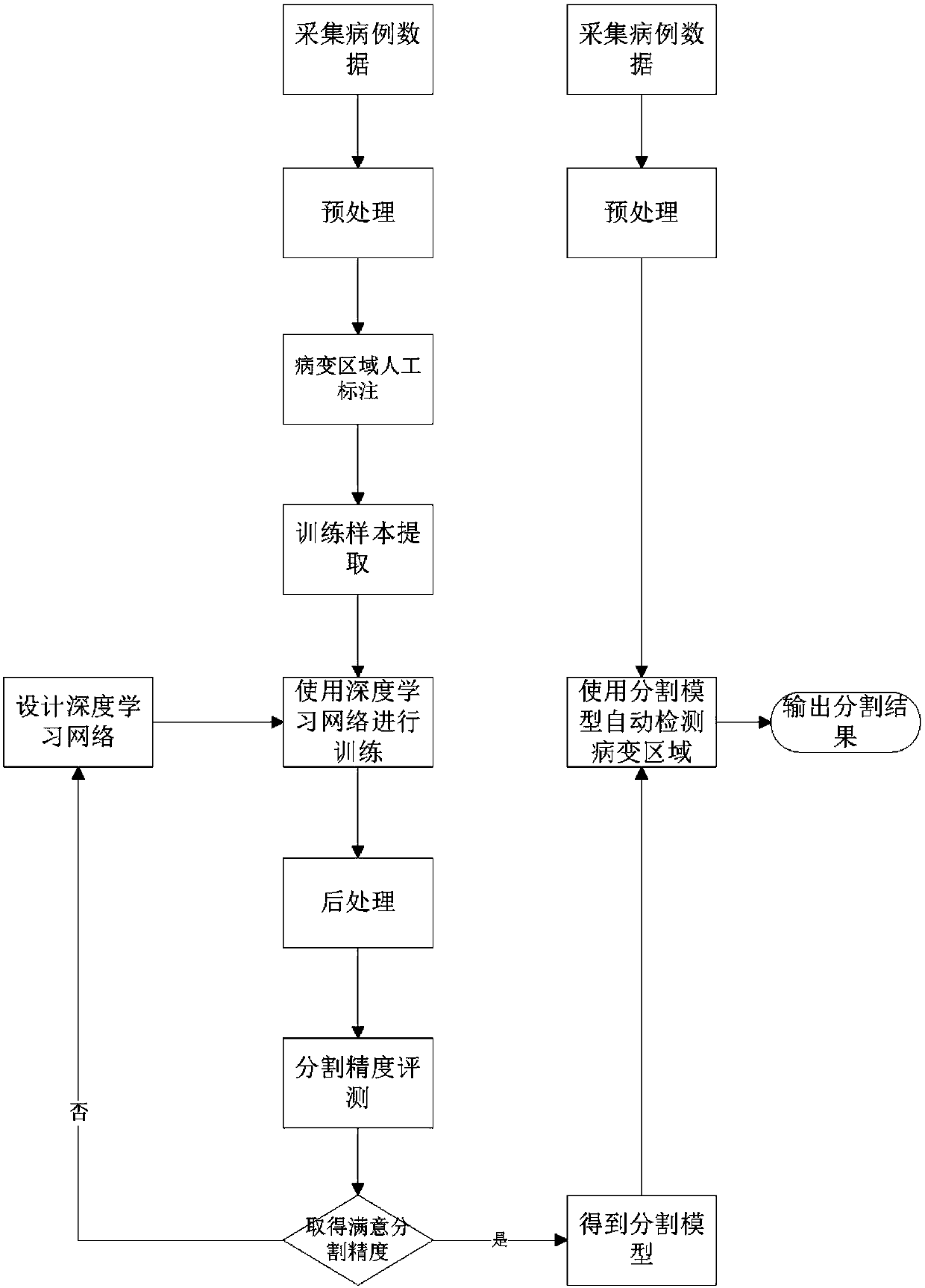
[0022] Such as figure 1 As shown, the automatic segmentation method of lesion area based on deep learning of the present invention comprises the following steps:

[0023] S1) Collect multiple case data, and then perform standardized preprocessing on the medical images of the specific modality of the lesion. Specifically, a number of cases with a certain type of disease are found, medical images of specific modalities of...


PUM


Abstract

The invention relates to an automatic segmentation method of pathology area based on deep learning, comprising the steps of S1 collecting multiple case data and conducting standardized preprocessing on medical images of a pathology position specific modal; S2 conducting edge mark on the pathology area layer by layer by a medical science audio visual technician, and using the marks as real data; S3conducting extraction of the training sample, wherein a plurality of voxels are extracted at random from voxels in the pathology area and within a certain distance outside of the pathology area, andimage blocks having fixed size are used as the training sample for the next step with the voxels being the center; S4 establishing a deep learning nerve network, and conducting training on the positive and negative samples of the above cases; conducting post-processing and segmentation precision assessment, and obtaining a segmentation model after the satisfied segmentation precision is obtained;S5 collecting medical science images of the same modal at the same position, and conducting standardized preprocessing for cases to be diagnosed; S6 automatically detecting the pathology area by meansof the segmentation model, and outputting the segmentation result.

Description

technical field [0001] The invention relates to the field of medical image processing, in particular to a method for automatically segmenting lesion regions based on deep learning. Background technique [0002] At present, the processing of medical images is still dominated by manual methods. Areas that may have larger lesions are selected in two-dimensional or three-dimensional images, and medical imaging specialists manually describe them on the two-dimensional images, and doctors then make diagnoses based on manual labeling results. and corresponding treatment. Some medical institutions have begun to introduce some auxiliary diagnostic equipment to find suspected lesion areas based on shape or abnormality detection, and further confirmation and edge delineation are performed manually. [0003] For physical lesions of the human body such as tumors, a variety of medical instruments can be used to examine the human body and generate human body images of different modalities...

Claims


Application Information

Patent Timeline
Patent Type & Authority Applications(China)
IPC IPC(8): G06T7/11G06N3/04G06N3/08G06T5/00
CPCG06N3/08G06T5/002G06T7/11G06T2207/30096G06T2207/20081G06T2207/20084G06N3/045
Inventor 冯翱马宗庆
Owner CHENGDU UNIV OF INFORMATION TECH
Why Eureka
Try Eureka