Automatic classification method for breast tumor ultrasound images

An ultrasound image and automatic classification technology, which is applied to instruments, character and pattern recognition, computer parts, etc., can solve the problems of low automatic classification accuracy of breast tumor ultrasound images, and achieve the effect of improving the recognition accuracy

Inactive Publication Date: 2018-03-02
高东平
View PDF0 Cites 10 Cited by
  • Summary
  • Abstract
  • Description
  • Claims
  • Application Information

AI Technical Summary

Problems solved by technology

[0005] The purpose of the present invention is to solve the problem of low automatic classification accuracy of ultrasound images of breast tumors, and closely combine the quantitative features of ultrasound images of breast tumors to quantify the histogram features, color features, contour features, bound

Method used


Image

  • Automatic classification method for breast tumor ultrasound images
  • Automatic classification method for breast tumor ultrasound images
  • Automatic classification method for breast tumor ultrasound images

Examples


Embodiment Construction

[0030] The specific implementation manners of the present invention will be described in further detail below in conjunction with the accompanying drawings and examples.

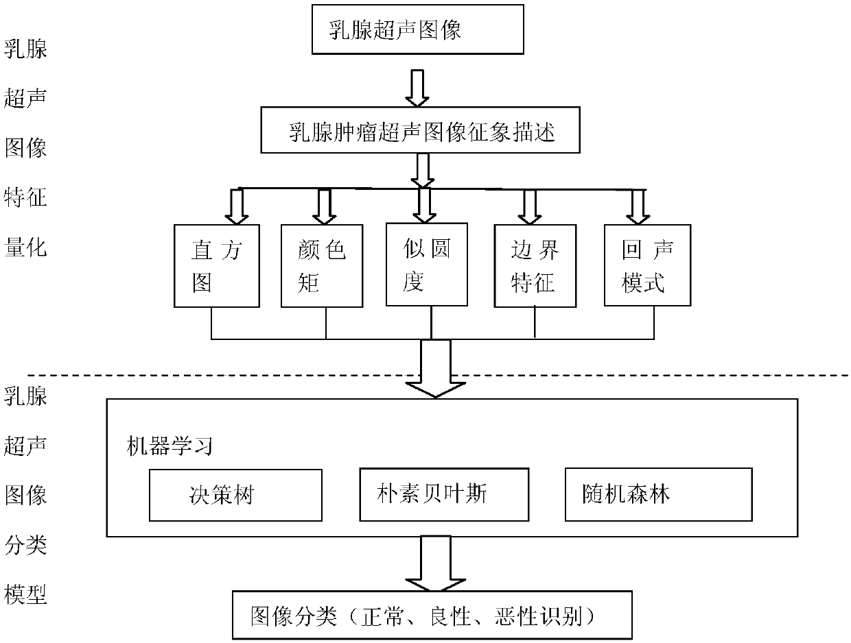
[0031] figure 1 Be the flow chart of the inventive method, carry out corpus pretreatment according to step 1, realize steps are as follows:

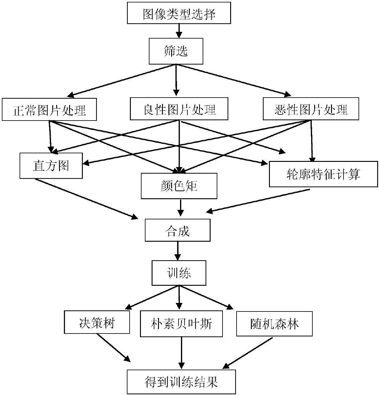
[0032] Step 1. Select the image type: divide the images into normal images, benign tumor images and malignant tumor images according to certain rules according to the source, and put them in different folders;

[0033] Step 2. Initialize the directories of normal images, benign tumor images, and malignant tumor images, initialize the test sample matrix, initialize the total number of files, and the number of currently processed files;

[0034] Step 3. Process normal pictures, benign tumor pictures, and malignant tumor pictures, and extract various features under each folder, including the histogram feature of the picture, color features, and contour features extracted...


PUM


Abstract

The invention relates to an automatic classification method for breast tumor ultrasound images, and belongs to the technical field of applications of artificial intelligence. According to the invention, quantitative features of the breast tumor ultrasound images are tightly combined, histogram features, color features, profile features, edge features and echo features in the breast tumor ultrasound images are quantified, a decision-making tree, a Naive Bayesian method and a random forest method are selected to serve as machine learning methods, and a decision-making tree, Naive Bayesian and random forest weighted fusion multi-dimensional classification method is proposed to perform recognition on the breast images. The automatic classification method can effectively improve the automatic classification accuracy of the breast tumor ultrasound images.

Description

technical field [0001] The invention relates to an automatic classification method for ultrasound images of breast tumors, belonging to the technical field of artificial intelligence applications. Background technique [0002] At present, the clinical analysis of medical images is mainly completed through the qualitative evaluation of images by doctors, and there is a lack of quantitative measurement of image features. Differences in people's visual perception, experience accumulation, and use of different characteristics and diagnostic criteria lead to differences in the diagnostic results of different doctors. Through computer methods, image features can be extracted and analyzed objectively and quantitatively to solve the limitations of human vision. Image-based computer-aided diagnosis was developed out of this need. [0003] For the automatic classification of breast ultrasound images, from the perspective of recognition methods, the image recognition methods mentione...

Claims


Application Information

Patent Timeline
IPC IPC(8): G06K9/46G06K9/62
CPCG06V10/50G06V10/44G06V10/56G06V2201/032G06F18/24155G06F18/24G06F18/214
Inventor 高东平
Owner 高东平
Why Eureka
Try Eureka