Method and device for enhancing CT image sequence

A CT image and image enhancement technology, which is applied in image enhancement, image analysis, image data processing, etc., can solve problems such as the influence of disease diagnosis accuracy and CT image blur

Active Publication Date: 2018-03-30
HANGZHOU ZHUOJIAN INFORMATION TECH CO LTD
View PDF5 Cites 8 Cited by
  • Summary
  • Abstract
  • Description
  • Claims
  • Application Information

AI Technical Summary

Problems solved by technology

[0004] The present invention provides a CT image sequence enhancement method and device to overcome the problem in the prior art that each CT image in the CT image sequence obtained by thick-slice scanning is relatively blurred, which will have a great impact on the accuracy of disease diagnosis

Method used


Image

  • Method and device for enhancing CT image sequence
  • Method and device for enhancing CT image sequence

Examples


Embodiment Construction

[0023] The specific implementation manners of the present invention will be further described in detail below in conjunction with the accompanying drawings and embodiments. The following examples are used to illustrate the present invention, but are not intended to limit the scope of the present invention.

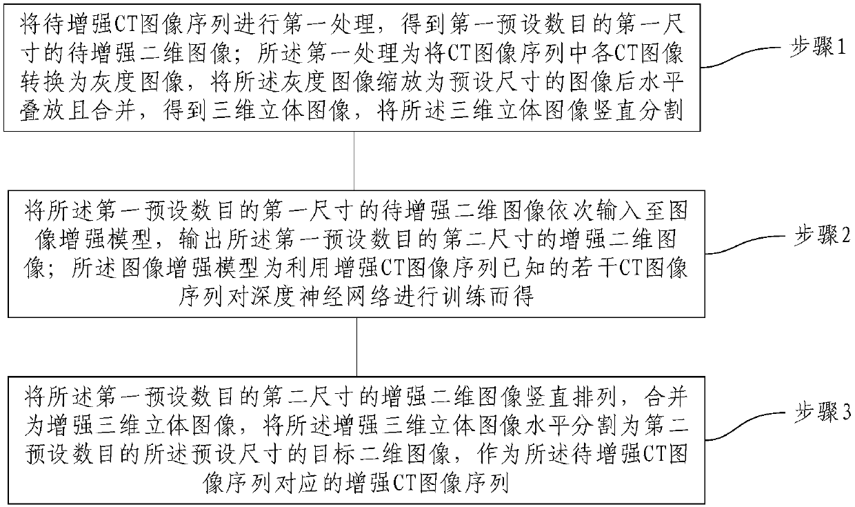
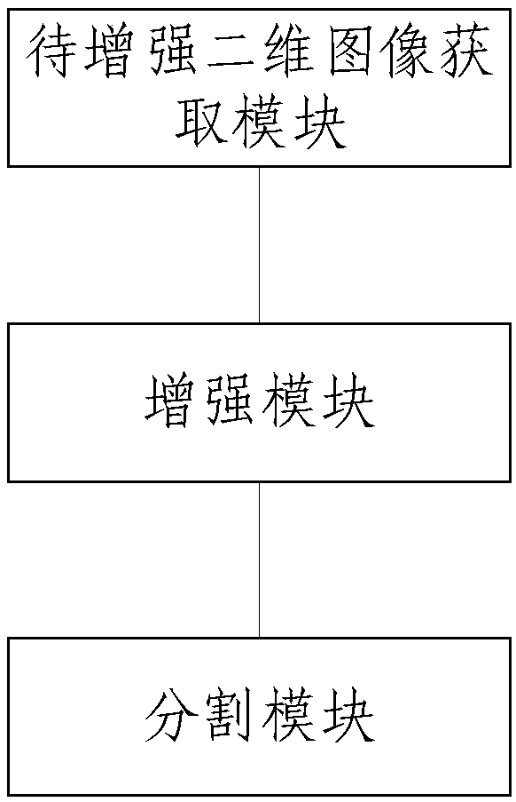
[0024] Such as figure 1 As shown, according to the first aspect of the present invention, a method for enhancing a CT image sequence is provided. The method includes: Step 1: Preprocessing the CT image sequence to be enhanced to obtain a first preset number of images of a first size to be enhanced. Enhancing the two-dimensional image; the preprocessing is converting each CT image in the CT image sequence into a grayscale image, scaling the grayscale image to an image of a preset size and then stacking and merging it horizontally to obtain a three-dimensional stereoscopic image. The three-dimensional stereoscopic image is vertically divided; step 2, input the first preset ...


PUM


Abstract

The invention provides a method and a device for enhancing a CT image sequence. The method includes the following steps: converting each CT image in a to-be-enhanced CT image sequence into a gray image, scaling the gray images into images with a preset size, horizontally stacking and combining the images to get a 3D stereoscopic image, and vertically segmenting the 3D stereoscopic image to get a first preset number of to-be-enhanced 2D images with a first size; inputting the first preset number of to-be-enhanced 2D images with the first size to an image enhancement model in turn, and outputting a first preset number of enhanced 2D images with a second size; and vertically arranging and combining the first preset number of enhanced 2D images with the second size into an enhanced 3D stereoscopic image, horizontally segmenting the enhanced 3D stereoscopic image into a second preset number of target 2D images with the preset size, and taking the second preset number of target 2D images with the preset size as an enhanced CT image sequence corresponding to the to-be-enhanced CT image sequence. Enhancement of the to-be-enhanced CT image sequence is realized.

Description

technical field [0001] The present invention relates to the technical field of image enhancement, and more particularly, to a method and device for enhancing a CT image sequence. Background technique [0002] CT (Computed Tomography), that is, electronic computer tomography, uses precisely collimated X-ray beams, γ-rays, ultrasound, etc., together with highly sensitive detectors, to perform cross-sectional scans one after another around a certain part of the human body. [0003] Scanning can be divided into thin layer scanning and thick layer scanning. Each CT image in the CT image sequence obtained by thin-layer scanning can well reflect the internal structure of the subject's human body, and has great reference value for the diagnosis of diseases. However, the X-ray dose of thin-layer scanning is relatively large, which is likely to have an impact on human health. Each CT image in the CT image sequence obtained by thick-slice scanning is relatively blurred, which will ha...

Claims


Application Information

Patent Timeline
Patent Type & Authority Applications(China)
IPC IPC(8): G06T5/00G06N3/04
CPCG06T5/001G06T2207/20081G06T2207/10012G06T2207/10081G06N3/045
Inventor 李玉杰刘立
Owner HANGZHOU ZHUOJIAN INFORMATION TECH CO LTD
Why Eureka
Try Eureka