Mass Spectrometry Probe for Glutamate Receptor and Detection Method of Spatial Distribution in Brain Tissue

A glutamate receptor and spatial distribution technology, applied in the field of mass spectrometry imaging, can solve the problems of poor quantitative relationship of absolute mass spectrometry signals, poor quantitative relationship of absolute mass spectrometry signals, and inability to solve the problem of quantitative comparison of different molecules

Active Publication Date: 2020-10-16
HUAZHONG NORMAL UNIV
View PDF8 Cites 0 Cited by
  • Summary
  • Abstract
  • Description
  • Claims
  • Application Information

AI Technical Summary

Problems solved by technology

However, different molecules have different vaporization and ionization capabilities, so the quantitative relationship between their absolute mass spectrometry signal intensity and content is poor. Realizing the quantitative analysis of different molecules by mass spectrometry has always been a difficult problem for mass spectrometry
[0003] Conventional MALDI (Matrix Assisted Laser Desorption Ionization) is to perform mass spectrometry through the protonation or deprotonation of sample molecules, or the formation of complexes with metal ions to become positive ions or negative ions. Due to the different vaporization and ionization capabilities of various molecules, The quantitative relationship between the absolute mass spectrum signal and the content is not good
The traditional stable isotope labeling method can only solve the problem of quantitative analysis of the same molecule in different samples, but cannot solve the problem of quantitative comparison between different molecules, and the quantification of different molecules is of great significance to full-scan mass spectrometry imaging

Method used


Image

  • Mass Spectrometry Probe for Glutamate Receptor and Detection Method of Spatial Distribution in Brain Tissue
  • Mass Spectrometry Probe for Glutamate Receptor and Detection Method of Spatial Distribution in Brain Tissue
  • Mass Spectrometry Probe for Glutamate Receptor and Detection Method of Spatial Distribution in Brain Tissue

Examples


Embodiment 1

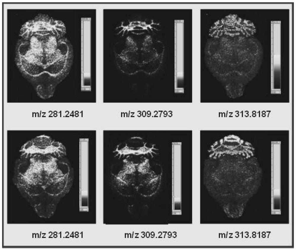
[0040] For high-resolution molecular imaging of other endogenous substances in the mouse brain that has not been injected with mass spectrometry probes, the steps are as follows:

[0041] 1) Preparation of image acquisition film: (Bi 2 o 3 ) 0.07 (CoO) 0.03 (ZnO) 0.9 The nano-semiconductor material is placed on a conductive aluminum or copper strip, and an image acquisition film is made under a pressure of 10MPa. The prepared image acquisition film is attached and fixed to the metal sample target to avoid the generation of air bubbles;

[0042] 2) Prepare anesthesia solution and prepare tissue cleaning solution: use analytically pure diethyl ether as an anesthetic, weigh analytically pure NaCl, dissolve it in pure water, and obtain physiological saline, which is used as a cleaning solution for rat brain tissue blood stains, and refrigerate at -4°C for later use;

[0043] 3) After anesthetizing the mouse with the anesthetic prepared in step 2), the brain was removed, and th...

Embodiment 2

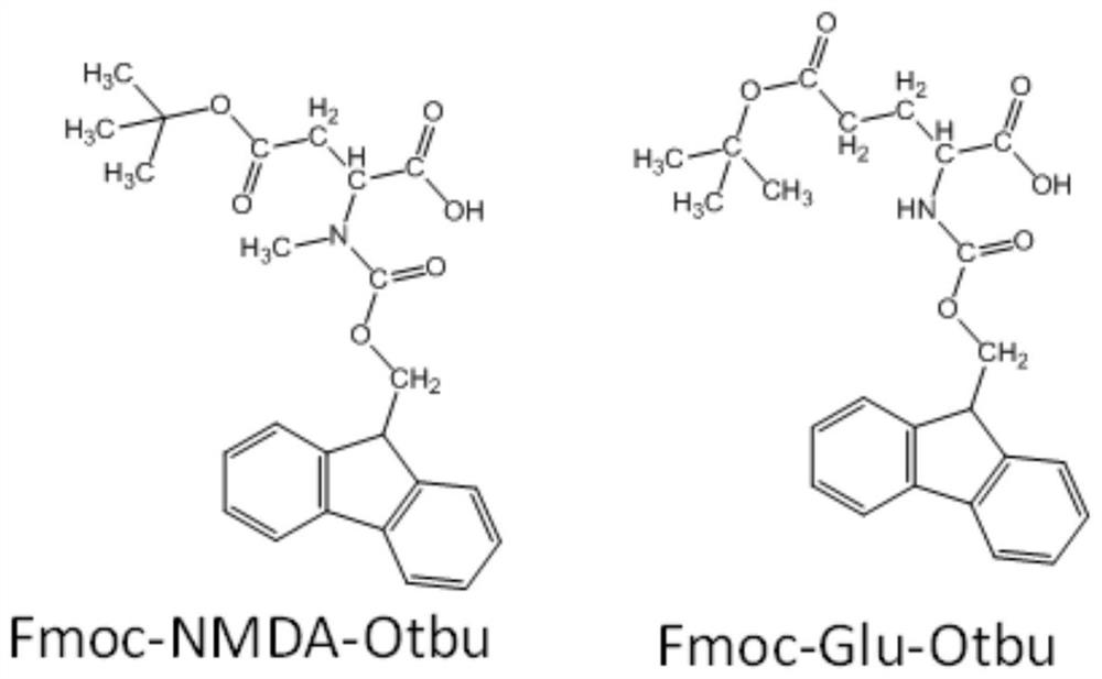
[0051] Spatialization of mass spectrometry probes (tert-butyl-protected glutamate conjugated to fluorenylmethoxycarbonyl group and tert-butyl-protected N-methyl-aspartic acid conjugated to fluorenylmethoxycarbonyl group) in the rat brain For distribution imaging, the operation steps are as follows:

[0052] 1) Prepare the probe molecule solution: Weigh the tert-butyl-protected glutamic acid combined with the fluorenyl moxycarbonyl group and the tert-butyl-protected N-methyl-aspartic acid combined with the fluorenyl moxycarbonyl group , dissolved in 50Mm NH 4 HCO 3 In normal saline, its concentration is 75mM;

[0053] 2) Place the solution obtained in step 1) on the surface of the image acquisition film with a pipette. After the solvent evaporates, put the sample target into the instrument for analysis, and adjust the voltage of the sample target, hexapod, extraction plate, and slit so that The voltage difference between the sample target and the slit is 20 volts;

[0054] ...

Embodiment 3

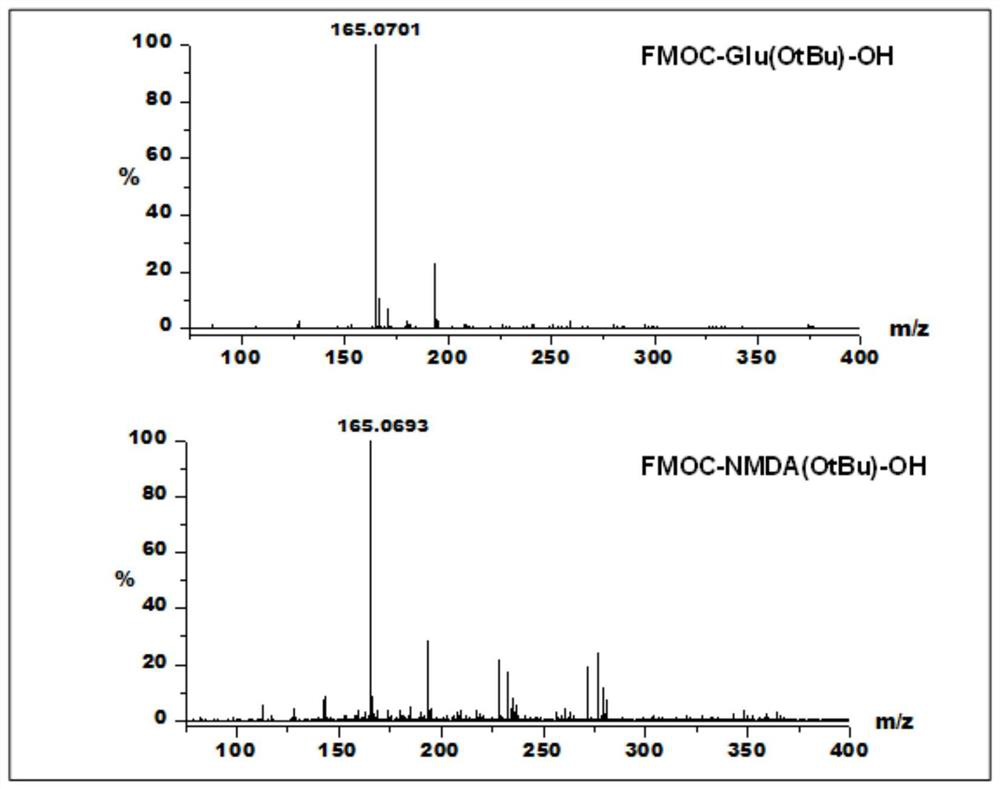
[0060] The spatial distribution imaging of mass spectrometry probe molecules (glutamic acid combined with fluorenyl moxycarbonyl group and N-methyl-aspartic acid combined with fluorenyl moxycarbonyl group) in the mouse brain, the operation steps are as follows :

[0061] 1) Prepare the probe molecule solution: Weigh the glutamic acid combined with the fluorenyl moxycarbonyl group and the N-methyl-aspartic acid combined with the fluorenyl methaneoxycarbonyl group, and dissolve them in 50Mm NH 4 HCO 3 In normal saline, its concentration is 75mM;

[0062] 2) Injecting the mass spectrometer probe solution obtained in step 1) into the mouse intravenously;

[0063] 3) The mouse brain obtained in step 2) was taken out and cleaned, and then cut into 20 micron thick tissue sections with a cryostat;

[0064] 4) Attach the tissue slice obtained in step 3) to the surface of the image acquisition film, put the sample target into the instrument for analysis, adjust the voltage of the sam...


PUM


Abstract

The invention relates to a mass spectrometry probe for a glutamate receptor as well as a method for detecting the spatial distribution rule of the mass spectrometry probe in a brain tissue. The mass spectrometry probe of the glutamate receptor consists of two parts, wherein one part is glutamic acid, N-methyl-aspartic acid, tertiary butyl protected glutamic acid or N-methyl-glutamic acid, and theother part is a fluorenoxycarbonyl group. The photoelectron tunnel effect of a semiconductor nanometer material generated under laser irradiation and the photoelectron capture capability of an electrophilic group in the mass spectrometry probe molecule are utilized, the light-generated electron is captured by an electron-deficient atom in the electrophilic group and breakage of the adjacent alpha-position chemical bond is initiated, different mass spectrometry probe molecules generate the same fragment ions of m / z 165.0702 Da, and quantitative comparison of the spatial distribution of different probe molecules can be realized by measuring the signal intensity; and the mass spectrometry probe molecule can be bound with a specific active cavity of the glutamate receptor, has the capability of penetrating through blood brain barrier and can provide a new thought and a new technology for medicine screening.

Description

technical field [0001] The invention belongs to the field of mass spectrometry imaging, and in particular relates to a glutamate receptor mass spectrometry probe and a high-resolution molecular imaging method. Background technique [0002] Mass spectrometry imaging is a new molecular imaging method using ion full scan and image reconstruction technology, which can be applied to determine the distribution and metabolism of drugs in different tissues. Since mass spectrometry is based on the determination of gaseous charged ions, neutral molecules will first be converted into gaseous charged ions for mass spectrometry. However, the vaporization and ionization capabilities of different molecules are different, so the quantitative relationship between the absolute mass spectrometry signal intensity and the content is poor. Realizing the quantitative analysis of different molecules by mass spectrometry has always been a difficult problem for mass spectrometry. [0003] Convention...

Claims


Application Information

Patent Timeline
Patent Type & Authority Patents(China)
IPC IPC(8): G01N27/64
CPCG01N27/64
Inventor 钟鸿英姜若葳张娟
Owner HUAZHONG NORMAL UNIV
Why Eureka
Try Eureka