Method, device and implementation device for detecting liver occupying infection focus area

A region detection and lesion technology, applied in the field of medical imaging, can solve the problem of low accuracy of the identification method of liver space-occupying lesions, and achieve the effect of improving the accuracy and increasing the image difference.

Inactive Publication Date: 2018-06-15
BEIJING PEREDOC TECH CO LTD
View PDF5 Cites 11 Cited by
  • Summary
  • Abstract
  • Description
  • Claims
  • Application Information

AI Technical Summary

Problems solved by technology

[0003] Enhanced CT scanning can more clearly highlight the characteristics of liver cancer areas, but limited by equipment, experience, objective recognition ability of the hum

Method used


Image

  • Method, device and implementation device for detecting liver occupying infection focus area
  • Method, device and implementation device for detecting liver occupying infection focus area
  • Method, device and implementation device for detecting liver occupying infection focus area

Examples


Embodiment Construction

[0025] In order to make the purpose, technical solutions and advantages of the embodiments of the present invention clearer, the technical solutions of the present invention will be clearly and completely described below in conjunction with the accompanying drawings. Obviously, the described embodiments are part of the embodiments of the present invention, not all of them. the embodiment. Based on the embodiments of the present invention, all other embodiments obtained by persons of ordinary skill in the art without making creative efforts belong to the protection scope of the present invention.
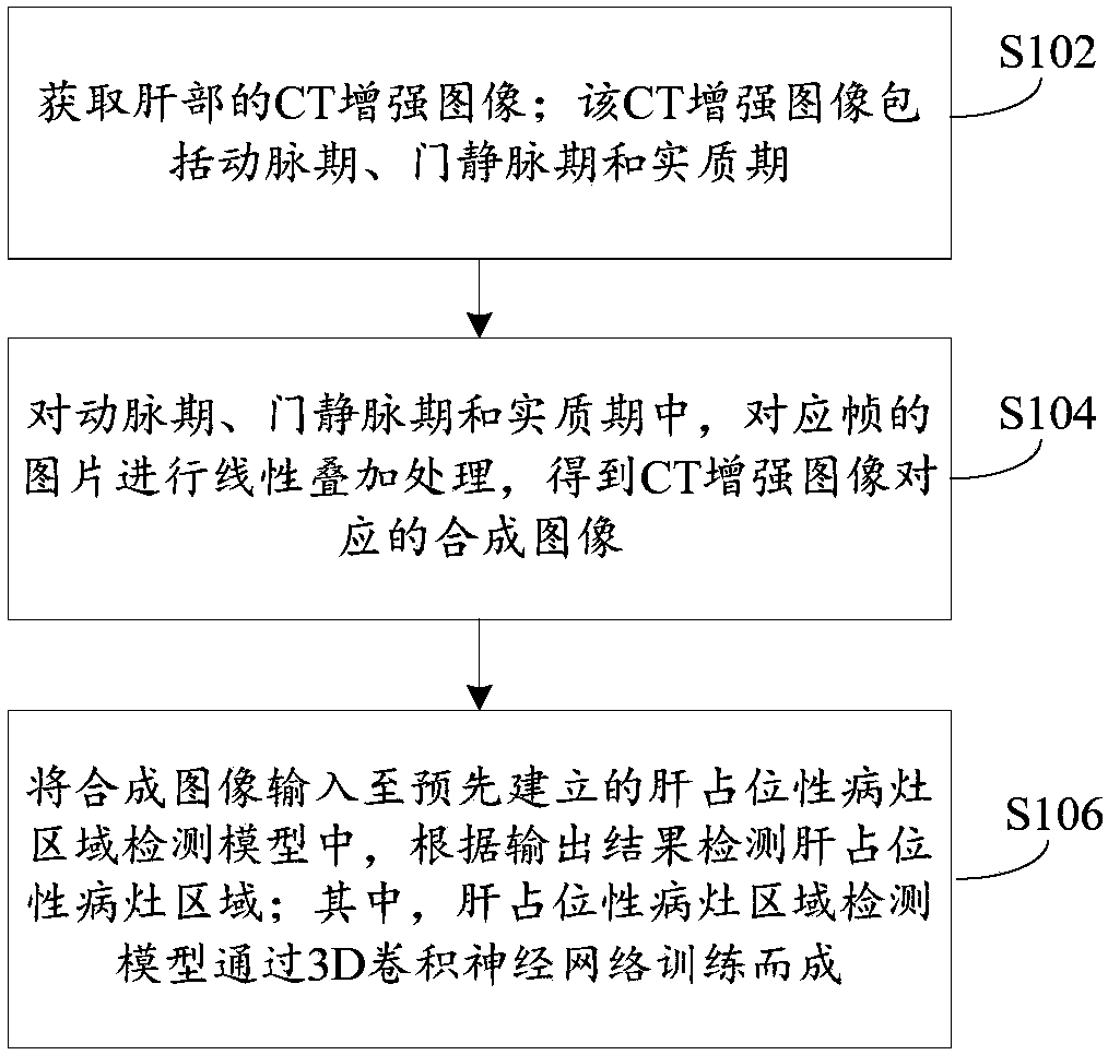
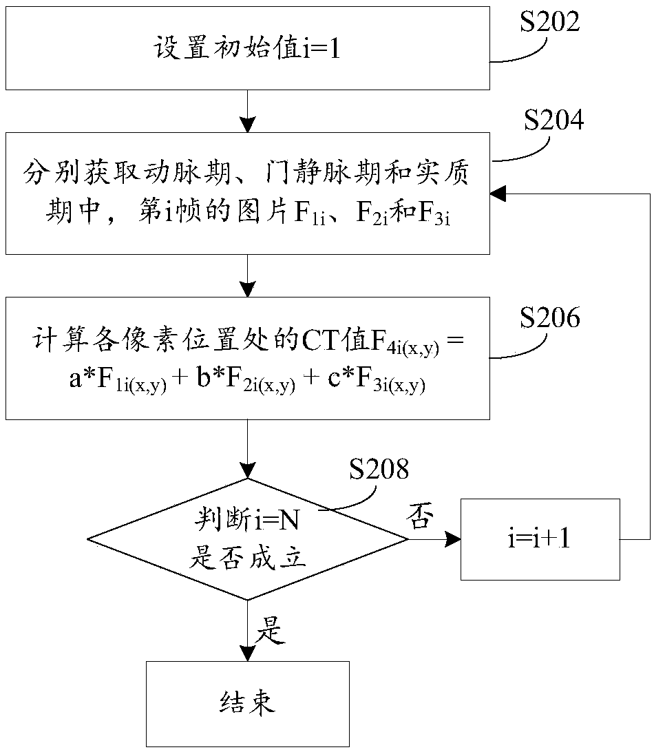
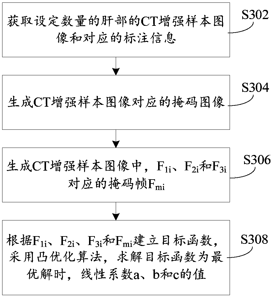
[0026] Usually, the cancerous area of ​​the liver or other hepatic space-occupying lesion area shows a lower density than the surrounding normal liver tissue on ordinary CT scan, which can show the size and shape of the lesion area. However, there are a small number of liver cancers whose density is not much different from that of the surrounding liver tissue, and ordinary scans cann...


PUM


Abstract

The invention provides a method, a device and an implementation device for detecting a liver occupying infection focus area. The method comprises the following steps: a step of acquiring CT enhanced images of a liver, wherein the CT enhanced images comprise arterial phase images, portal vein phase images and parenchymal phase images; the images for each phase include a plurality frames of image; in the arterial phase, the portal vein phase and the parenchymal phase, the images of corresponding frames are linearly superimposed to obtain composite images corresponding to the CT enhanced images;the composite images are input to a pre-established liver occupying infection focus area detection model, the liver occupying infection focus area is detected according to an output result, and the liver occupying infection focus area detection model is trained via a 3D convolutional neural network. According to the method, the three-phase images in the CT enhanced images are linearly superposed,image difference between the infection focus area and a normal area can be increased, the model which is trained via the3D convolutional neural network is used for detecting the liver occupying infection focus area, and accuracy of a liver occupying infection focus area identification mode can be improved.

Description

technical field [0001] The present invention relates to the technical field of medical imaging, in particular to a detection method, device and implementation device for a liver space-occupying lesion area. Background technique [0002] Enhanced CT scanning can make up for the deficiency of ordinary CT scanning by observing the dynamic changes of liver cancer, surrounding liver and blood vessel density. For example, in the early stage (arterial phase) after injection of contrast medium, liver cancer is significantly enhanced and densely supplied by the hepatic artery, and the surrounding liver tissue mainly has portal vein blood supply that is not significantly enhanced and relatively low in density; Stage), the liver cancer no longer enhanced, the density decreased, and the surrounding liver tissue enhanced significantly, and the density increased. Therefore, liver cancer can be displayed more clearly by scanning at different times after contrast agent injection. [0003]...

Claims


Application Information

Patent Timeline
IPC IPC(8): G06T7/00G06N3/08G06T5/50
CPCG06T7/0012G06N3/08G06T5/50G06T2207/10081G06T2207/30056
Inventor 胡飞王方
Owner BEIJING PEREDOC TECH CO LTD
Why Eureka
Try Eureka