Identification method of abnormal cells in pathological sections based on multiscale expansion convolution

A technology for pathological slices and abnormal cells, applied in character and pattern recognition, acquisition/recognition of microscopic objects, instruments, etc., can solve the problem of no semantic segmentation model, achieve good test results, high accuracy and precision, and reduce the burden the effect of the workload

Inactive Publication Date: 2018-06-22
ZHEJIANG UNIV
View PDF4 Cites 23 Cited by
  • Summary
  • Abstract
  • Description
  • Claims
  • Application Information

AI Technical Summary

Problems solved by technology

For more complex medical images, there is...

Method used


Image

  • Identification method of abnormal cells in pathological sections based on multiscale expansion convolution
  • Identification method of abnormal cells in pathological sections based on multiscale expansion convolution
  • Identification method of abnormal cells in pathological sections based on multiscale expansion convolution

Examples


Embodiment Construction

[0050] In order to further understand the present invention, a method for identifying abnormal cells in pathological slices based on multi-scale dilation and convolution provided by the present invention will be described in detail below in conjunction with specific embodiments. The pathological slices in the specific embodiments of the present invention are based on gastric cancer pathological slices. Examples are described, but the present invention is not limited thereto, non-essential improvements and adjustments made by those skilled in the art under the core guiding ideology of the present invention still belong to the protection scope of the present invention.

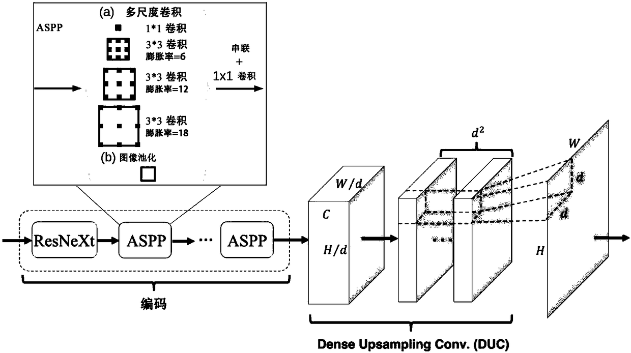
[0051] A method for identifying abnormal cells in pathological slices based on multi-scale dilated convolution, comprising the following steps:

[0052] Step 1: Preprocessing of input data

[0053] Firstly, the pathological image of gastric cancer is preprocessed, and the image is scaled to about *5 times, so th...


PUM


Abstract

The invention discloses an identification method of abnormal cells in pathological sections based on multiscale expansion convolution. The method comprises the steps of 1, preprocessing the electronically-scanned pathological sections to obtain training data; 2, setting up a multiscale expansion convolution division model, wherein a ResNeXt network serves as the basic framework of the model, ASPPmodules and DUC modules serve as the basic modules of the model, and number of the ASPP modules is 1 to 5; 3, adopting the training data obtained in step 1 to train the multiscale expansion convolution division model obtained in step 2, and adjusting parameters of the network according to a model predicting result and the overlap ratio of labels, so that the model is converged and the abnormal cells in the pathological sections are identified. By means of the identification method, the accuracy rate of sifting the abnormal cells is high, the sifting precision of the abnormal cells is high, andthe F1 value serves as an index of a measuring algorithm, and can reach 98% or above.

Description

technical field [0001] The invention belongs to the field of artificial intelligence, and in particular relates to a method for identifying abnormal cells in pathological slices. Background technique [0002] Abnormal cells in traditional pathological slides are screened manually: under the microscope, professional pathologists scan the entire slice with the naked eye through the movement of the slice to find whether there are abnormal cells in the entire slice. As the reading time increases, the error rate also increases. [0003] With the continuous development of technology, the identification of abnormal cells in pathological slides can be screened with the help of computers. Image semantic segmentation (Semantic Segmentation) is an important research direction of computer vision, and its task is to classify each pixel of a single image through computer algorithms. Semantic segmentation tasks have important applications in scenarios such as autonomous driving and objec...

Claims


Application Information

Patent Timeline
IPC IPC(8): G06K9/00G06K9/62
CPCG06V20/69G06F18/214
Inventor 吴健王彦杰王文哲刘雪晨吴边陈为吴福理吴朝晖
Owner ZHEJIANG UNIV
Why Eureka
Try Eureka