
Cervical cancer TCT digital section data analysis method based on ResNet

A technology of digital slicing and data analysis, applied in character and pattern recognition, instruments, computer parts, etc., to reduce costs, improve recognition efficiency, and prevent interference from external factors

Active Publication Date: 2018-07-27
NANJING ILUVATAR COREX TECH CO LTD (DBA ILUVATAR COREX INC NANJING)
View PDF7 Cites 21 Cited by
  • Summary
  • Abstract
  • Description
  • Claims
  • Application Information

AI Technical Summary

Problems solved by technology

At present, there is no unified process for cervical cancer screening in China. Some hospitals use TCT+HPV combined screening, while others continue to use simple cervical smear examination. used to shunt

Method used


Image

  • Cervical cancer TCT digital section data analysis method based on ResNet

Examples


Embodiment Construction

[0027] The following is based on figure 1 The specific embodiment of the present invention is further described:

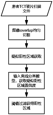
[0028] see figure 1 , a ResNet-based cervical cancer TCT digital slice data analysis method, comprising the following steps:

[0029] (1) Obtain the positive area in the cervical TCT digital slice image, where the positive area in the cervical TCT digital slice image is marked by a doctor, and train the autoencoder based on the obtained positive area samples to obtain the trained autoencoder. The above-mentioned positive area is the lesion area, and the lesion area is marked by the doctor, and the lesion area generally includes high-grade lesion area, low-grade lesion area and / or suspected lesion area;

[0030] (2) Input the positive area obtained in step (1) into the trained autoencoder to obtain the positive features in the positive area, and use the positive features in multiple positive areas as samples to train the single-class SVM classifier, and get A tr...


PUM


Abstract

The invention discloses a cervical cancer TCT digital section data analysis method based on ResNet. The method comprises the steps of obtaining a TCT slide scanning image of a patient, and uniformly dividing the TCT slide scanning image to obtain multiple image blocks which are uniformly cut; inputting the image blocks to an automatic coder to extract features, further inputting the extracted features into a single-class SVM classifier, and extracting image blocks belonging to a positive region; preprocessing the extracted image blocks, inputting the processed image blocks to a trained ResNetclassification model, obtaining the lesion confidence coefficients of the image blocks, presetting confidence coefficient threshold values, and determining the image blocks of which the lesion confidence coefficients are higher than the confidence coefficient threshold values as the positive region. By means of the cervical cancer TCT digital section data analysis method based on ResNet, cervicalcancer TCT digital section image data is detected. Compared with a traditional cervical cancer detection method, the disclosed cervical cancer TCT digital section data analysis method can save the image medical diagnosis time and cost, and improve the diagnosis and treatment accuracy.

Description

technical field [0001] The invention belongs to the application of deep learning in the medical field, and in particular relates to a ResNet-based analysis method for TCT digital slice data of cervical cancer. Background technique [0002] With the emergence of convolutional neural networks and the perfection of deep neural networks, artificial intelligence computer vision based on deep learning has developed rapidly in recent years. Li Feifei, a tenured professor of computer science at Stanford University, once said that the level of artificial intelligence can now begin to affect the medical and health field. make a contribution. [0003] The biggest feature of artificial intelligence (AI) is fast learning. The artificial intelligence model based on deep learning has a deep neural network medical model with random parameters, and then trains the model with marked data, adjusts the model parameters after errors occur, and then assists with Medical knowledge, through a larg...

Claims


Application Information

Patent Timeline
Patent Type & Authority Applications(China)
IPC IPC(8): G06K9/62G06K9/34
CPCG06V10/267G06F18/2411
Inventor 吕艳洁黄沈乾李晶晶
Owner NANJING ILUVATAR COREX TECH CO LTD (DBA ILUVATAR COREX INC NANJING)
Learn More