Multi-directional-contour-based medical image segmentation method

A medical image, multi-directional technology, applied in the field of medical image segmentation based on multi-directional contours, can solve problems such as difficult to determine boundaries, large subjective influence, increased workload, etc., to improve modeling efficiency and accuracy, and gray-scale dependence Small, the effect of improving segmentation accuracy

Active Publication Date: 2018-09-04
深圳市一图智能科技有限公司
View PDF4 Cites 0 Cited by
  • Summary
  • Abstract
  • Description
  • Claims
  • Application Information

AI Technical Summary

Benefits of technology

The technical effect of this patented method described in this patents allows for efficient and precise tooling of medical imagery data that includes various forms such as X rays or CT scans (computerized tomography). By generating contour shapes simultaneously from all three dimensions, it becomes possible to create models accurately without depending upon how well an xray machine produces certain kinds of radiation. Additionally, the use of single directional contortions simplifies the process of constructing surfaces within 3D space while still maintaining good quality results. Overall, this technique provides improved precision and speed when processing large amounts of patient data compared to previous methods like filtered backprojection techniques.

Problems solved by technology

This patents describes various technical problem addressed during medical procedures involving identifying specific areas within organs like brain cancer (brain cancer). Current techniques involve manually sectioning through X ray scanning imagery for identification purposes but these processes require significant effort due to their complexity and human error potential. There also exist existing automated systems used for pathology analysis without considering any depth relationships among them, resulting in imprecise boundaries being marked onto computed tomography/magnetic resonance images.

Method used


Image

  • Multi-directional-contour-based medical image segmentation method
  • Multi-directional-contour-based medical image segmentation method
  • Multi-directional-contour-based medical image segmentation method

Examples


Embodiment Construction

[0020] The present invention will be described in more detail below in conjunction with the accompanying drawings and embodiments.

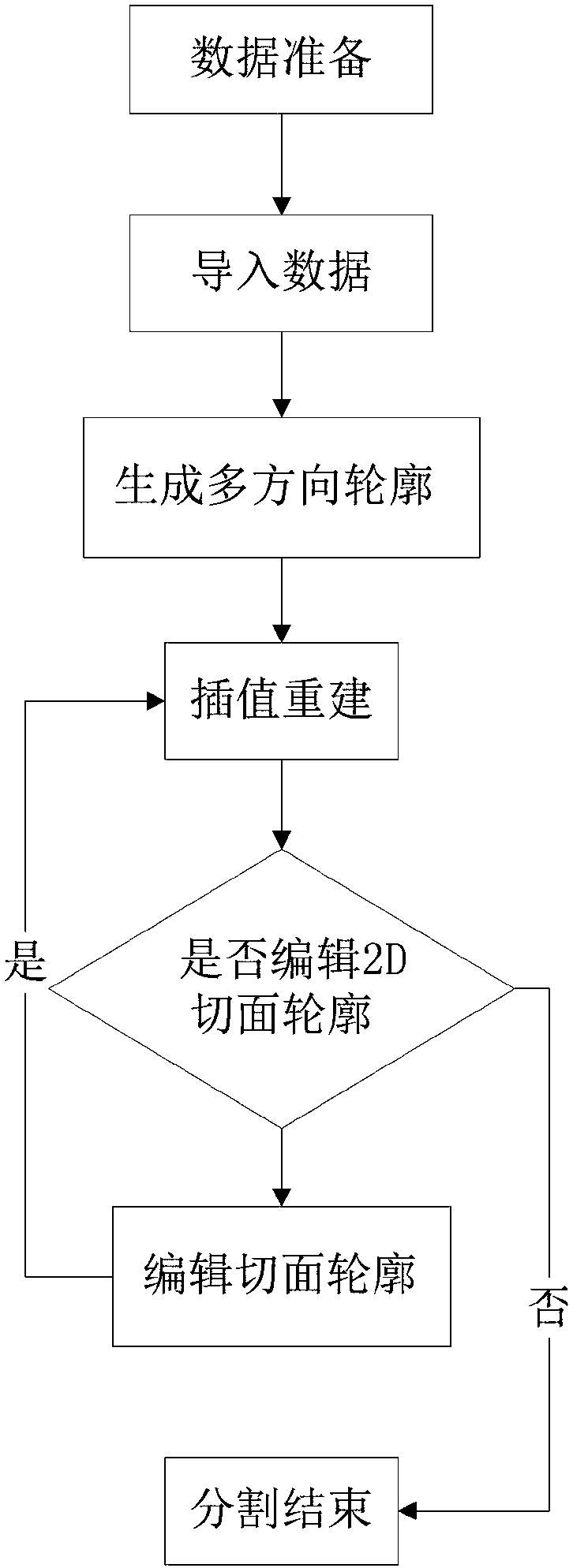
[0021] The invention discloses a medical image segmentation method based on multi-directional contours, please refer to figure 1 , which includes the following steps:

[0022] Step S1, data preparation, obtaining sequential thin-slice CT or MRI medical images;

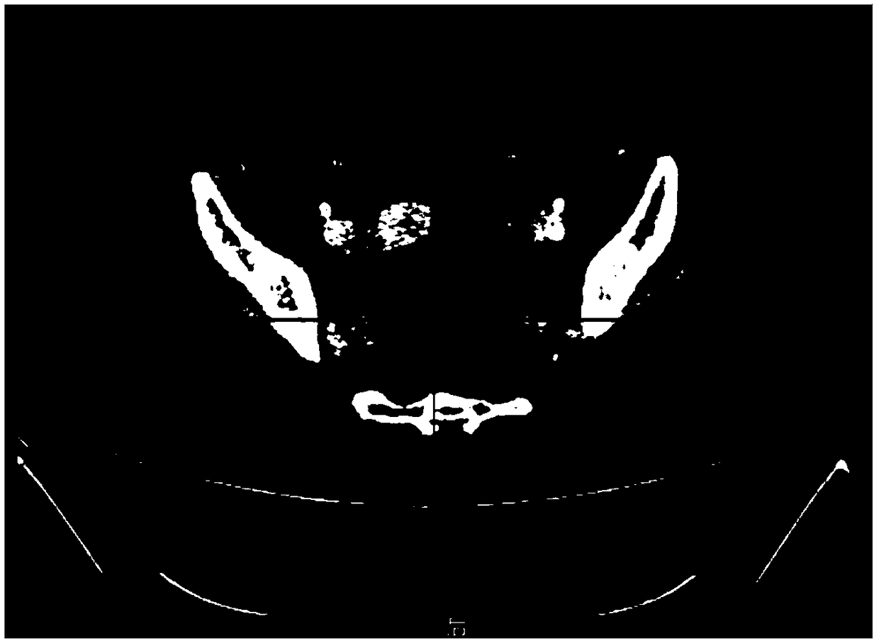
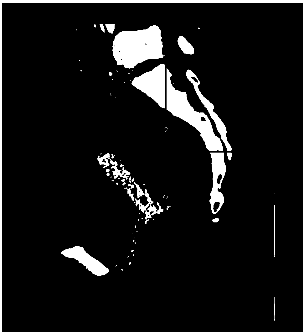
[0023] Step S2, import the sequence of medical images, perform MPR reconstruction on the images, and display the medical images in the window, the image cross-section, coronal plane and sagittal plane can be displayed in the display view, and any direction slice can be added and adjusted as needed for image display;

[0024] Step S3, select a multi-angle section for the segmentation object in the medical image, use the mouse to set a seed point inside the object to be segmented on any section, use the magic wand segmentation method to segment the approximate threshold area on the multi-...


PUM


Abstract

The invention discloses a multi-directional-contour-based medical image segmentation method. The method comprises: a medical image is inputted and MPR reconstruction is carried out; a multi-directional contour is generated based on a magic wand algorithm; interpolation reconstruction is carried out, an indication grid is generated, and a binary segmentation MASK is obtained; whether the binary segmentation MASK image is accurate is determined layer by layer; if so, a binary segmentation MASK image and a surface grid of a target region are outputted and a segmentation algorithm is ended; if not, a section contour is edited, a section not segmented inaccurately in the binary segmentation MASK image is selected, a contour line and key points of the binary segmentation MASK section image are extracted, local adjustment of the key points is carried out by using a mouse, and after ending of the adjustment, the above-mentioned steps are performed again and surface grid reconstruction is carried out again. According to the invention, contours of a to-be-segmented object at multiple sections in different direction are extracted instantly by one operation, shape constraints are established,curved surface reconstruction is carried out by using the interpolation reconstruction technology, and iterative segmentation contour optimization and network reconstruction are supported. Therefore,the modeling efficiency and precision are improved effectively.

Description


Claims


Application Information

Patent Timeline
Owner 深圳市一图智能科技有限公司
Why Eureka
Try Eureka