
A blood vessel segmentation method for fundus images

A fundus image and blood vessel technology, applied in the computer field, can solve the problems of missing information, inability to completely describe the characteristics of blood vessels, etc., and achieve the effect of accurate blood vessel segmentation

Active Publication Date: 2019-05-24
珠海全一科技有限公司
View PDF4 Cites 0 Cited by
  • Summary
  • Abstract
  • Description
  • Claims
  • Application Information

AI Technical Summary

Problems solved by technology

In the supervised scheme, the neural network model needs to extract image features layer by layer, and a lot of useful information is lost, resulting in the parameters learned by the neural network model not being able to fully describe the characteristics of blood vessels.

Method used


Image

  • A blood vessel segmentation method for fundus images
  • A blood vessel segmentation method for fundus images

Examples


Embodiment Construction

[0028] In order to make the purpose, technical solutions and advantages of the embodiments of the present invention clearer, the technical solutions in the embodiments of the present invention will be clearly and completely described below in conjunction with the drawings in the embodiments of the present invention. Obviously, the described embodiments It is a part of embodiments of the present invention, but not all embodiments. Based on the embodiments of the present invention, all other embodiments obtained by persons of ordinary skill in the art without making creative efforts belong to the protection scope of the present invention.

[0029] It should be noted that in this article, relative terms such as "first" and "second" are only used to distinguish one entity or operation from another entity or operation, and do not necessarily require or imply these No such actual relationship or order exists between entities or operations.

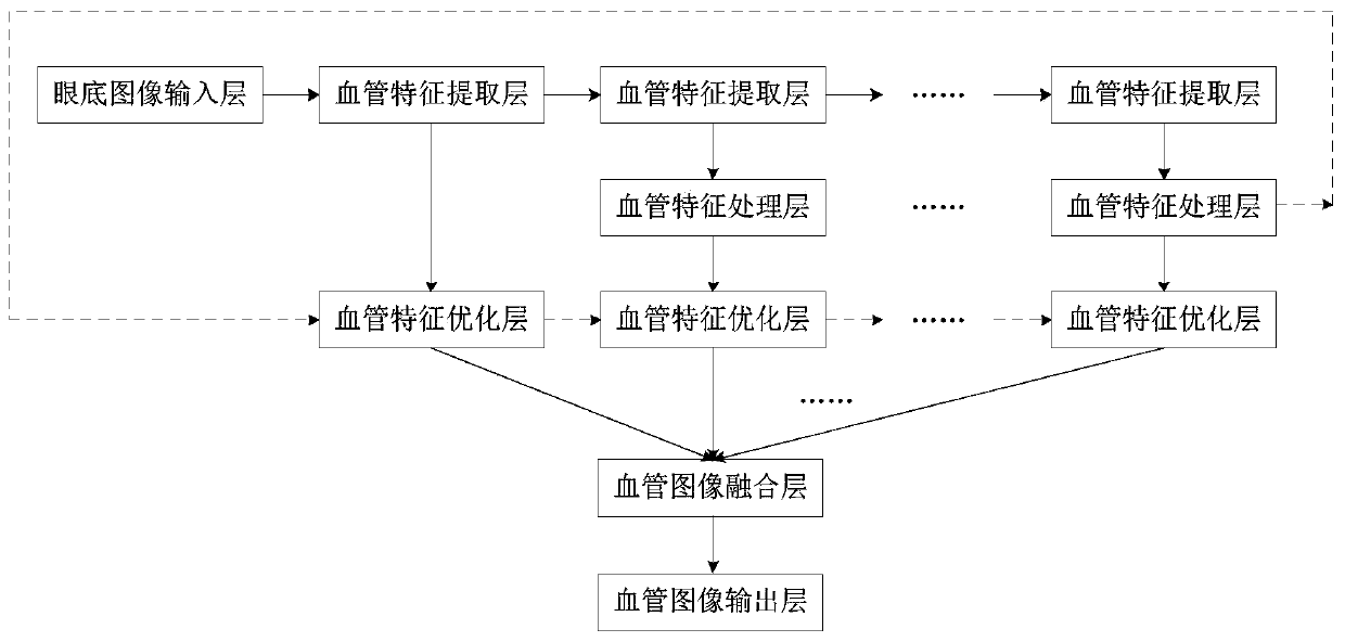
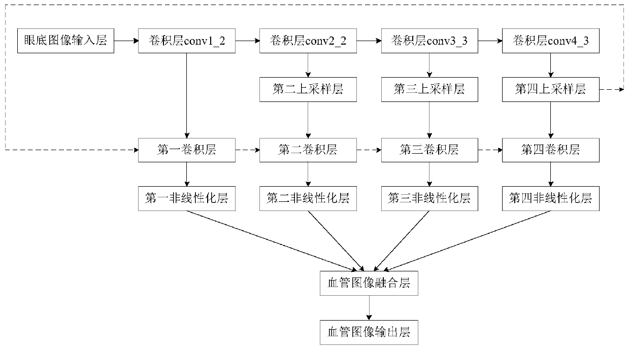
[0030] Such as figure 1 As shown, this ...


PUM


Abstract

The embodiment of the present invention relates to a method for segmenting blood vessels in a fundus image. Each blood vessel feature processing layer sends an up-sampled image obtained by this layer to a connected blood vessel feature optimization layer; the lowest blood vessel feature optimization layer also acquires the lowest blood vessel feature. The feature image of the extraction layer; the blood vessel feature processing layer of the highest layer sends the up-sampled image obtained by this layer to the blood vessel feature optimization layer of the lowest layer through the backward short connection; each blood vessel feature optimization layer sequentially acquires each image. Performing blood vessel feature extraction and non-linear processing to obtain a non-linear image corresponding to each image; each blood vessel feature optimization layer sends each acquired image to a higher blood vessel feature optimization layer through forward short connections. In the embodiment of the present invention, the high-level information is sent to the low-level through the backward short connection, and the low-level information is sent to the high-level through the forward short connection, so that the features of all levels are fully integrated, and the blood vessel segmentation is more accurate.

Description

technical field [0001] Embodiments of the present invention relate to the field of computer technology, and in particular to a method for segmenting blood vessels in fundus images. Background technique [0002] Retinal fundus image analysis helps ophthalmologists to deal with the diagnosis, screening and treatment of cardiovascular and ophthalmic diseases, such as macular degeneration, diabetic retinopathy, glaucoma, hypertension, etc. These diseases can lead to blindness if left untreated. Vessel segmentation is a fundamental step in retinal image analysis and helps localize diabetic retinopathy and foveal regions. However, in clinical practice, manual labeling of blood vessels in retinal images is time-consuming and requires a lot of experience. Therefore, automatic retinal vessel segmentation is necessary to reduce labeling time. [0003] The automatic segmentation schemes of retinal image vessels in recent decades can be divided into two categories: unsupervised schem...

Claims


Application Information

Patent Timeline
Patent Type & Authority Patents(China)
IPC IPC(8): G06K9/34G06N3/04
CPCG06V10/267G06N3/045
Inventor 季鑫
Owner 珠海全一科技有限公司
Learn More